

depth is greatest on the head and over the fins where scales
are absent. The thickness of epidermis and number of
mucous-secreting goblet cells in the epidermis varies
between species and location on the body. Their number
may increase during sexual maturation and spawning migration, and lymphocytes can also be present in the epidermis
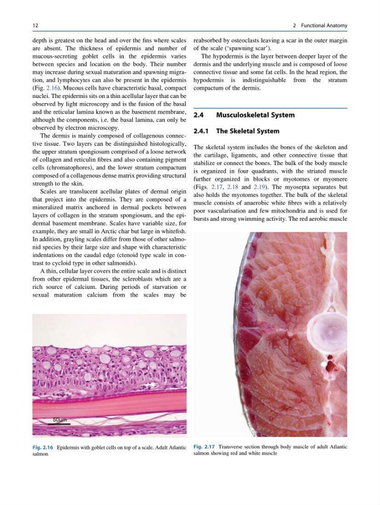
(Fig. 2.16). Mucous cells have characteristic basal, compact
nuclei. The epidermis sits on a thin acellular layer that can be
observed by light microscopy and is the fusion of the basal
and the reticular lamina known as the basement membrane,
although the components, i.e. the basal lamina, can only be
observed by electron microscopy.
The dermis is mainly composed of collagenous connective tissue. Two layers can be distinguished histologically,
the upper stratum spongiosum comprised of a loose network
of collagen and reticulin fibres and also containing pigment
cells (chromatophores), and the lower stratum compactum
composed of a collagenous dense matrix providing structural
strength to the skin.
Scales are translucent acellular plates of dermal origin
that project into the epidermis. They are composed of a
mineralized matrix anchored in dermal pockets between
layers of collagen in the stratum spongiosum, and the epidermal basement membrane. Scales have variable size, for
example, they are small in Arctic char but large in whitefish.
In addition, grayling scales differ from those of other salmonid species by their large size and shape with characteristic
indentations on the caudal edge (ctenoid type scale in contrast to cycloid type in other salmonids).
A thin, cellular layer covers the entire scale and is distinct
from other epidermal tissues, the scleroblasts which are a
rich source of calcium. During periods of starvation or
sexual maturation calcium from the scales may be
reabsorbed by osteoclasts leaving a scar in the outer margin
of the scale (‘spawning scar’).
The hypodermis is the layer between deeper layer of the
dermis and the underlying muscle and is composed of loose
connective tissue and some fat cells. In the head region, the
hypodermis is indistinguishable from the stratum
compactum of the dermis.
2.4
Musculoskeletal System
2.4.1 The Skeletal System
The skeletal system includes the bones of the skeleton and
the cartilage, ligaments, and other connective tissue that
stabilize or connect the bones. The bulk of the body muscle
is organized in four quadrants, with the striated muscle
further organized in blocks or myotomes or myomere
(Figs. 2.17, 2.18 and 2.19). The myosepta separates but
also holds the myotomes together. The bulk of the skeletal
muscle consists of anaerobic white fibres with a relatively
poor vascularisation and few mitochondria and is used for
bursts and strong swimming activity. The red aerobic muscle
Fig. 2.16 Epidermis with goblet cells on top of a scale. Adult Atlantic
salmon
Fig. 2.17 Transverse section through body muscle of adult Atlantic
salmon showing red and white muscle
12
2 Functional Anatomy
are absent. The thickness of epidermis and number of
mucous-secreting goblet cells in the epidermis varies
between species and location on the body. Their number
may increase during sexual maturation and spawning migration, and lymphocytes can also be present in the epidermis
(Fig. 2.16). Mucous cells have characteristic basal, compact
nuclei. The epidermis sits on a thin acellular layer that can be
observed by light microscopy and is the fusion of the basal
and the reticular lamina known as the basement membrane,
although the components, i.e. the basal lamina, can only be
observed by electron microscopy.
The dermis is mainly composed of collagenous connective tissue. Two layers can be distinguished histologically,
the upper stratum spongiosum comprised of a loose network
of collagen and reticulin fibres and also containing pigment
cells (chromatophores), and the lower stratum compactum
composed of a collagenous dense matrix providing structural
strength to the skin.
Scales are translucent acellular plates of dermal origin
that project into the epidermis. They are composed of a
mineralized matrix anchored in dermal pockets between
layers of collagen in the stratum spongiosum, and the epidermal basement membrane. Scales have variable size, for
example, they are small in Arctic char but large in whitefish.
In addition, grayling scales differ from those of other salmonid species by their large size and shape with characteristic
indentations on the caudal edge (ctenoid type scale in contrast to cycloid type in other salmonids).
A thin, cellular layer covers the entire scale and is distinct
from other epidermal tissues, the scleroblasts which are a
rich source of calcium. During periods of starvation or
sexual maturation calcium from the scales may be
reabsorbed by osteoclasts leaving a scar in the outer margin
of the scale (‘spawning scar’).
The hypodermis is the layer between deeper layer of the
dermis and the underlying muscle and is composed of loose
connective tissue and some fat cells. In the head region, the
hypodermis is indistinguishable from the stratum
compactum of the dermis.
2.4
Musculoskeletal System
2.4.1 The Skeletal System
The skeletal system includes the bones of the skeleton and
the cartilage, ligaments, and other connective tissue that
stabilize or connect the bones. The bulk of the body muscle
is organized in four quadrants, with the striated muscle
further organized in blocks or myotomes or myomere
(Figs. 2.17, 2.18 and 2.19). The myosepta separates but
also holds the myotomes together. The bulk of the skeletal
muscle consists of anaerobic white fibres with a relatively
poor vascularisation and few mitochondria and is used for
bursts and strong swimming activity. The red aerobic muscle
Fig. 2.16 Epidermis with goblet cells on top of a scale. Adult Atlantic
salmon
Fig. 2.17 Transverse section through body muscle of adult Atlantic
salmon showing red and white muscle
12
2 Functional Anatomy