

and the bulbus arteriosus, interposed by the conus arteriosus.
The former was considered to have been lost through evolution but recent work has clearly shown its presence and
fundamental role in the heart outflow tract, where it supports
the conus valves previously named bulbo-ventricular valves.
Deoxygenated blood from the cardinal and hepatic veins
flows into the thin-walled sinus venosus and passes through
the sino-atrial valve and then into the thin-walled spongy
atrium. Blood is drawn from the atrium via the atrio ventricular valves into the thick-walled and muscular ventricle. The
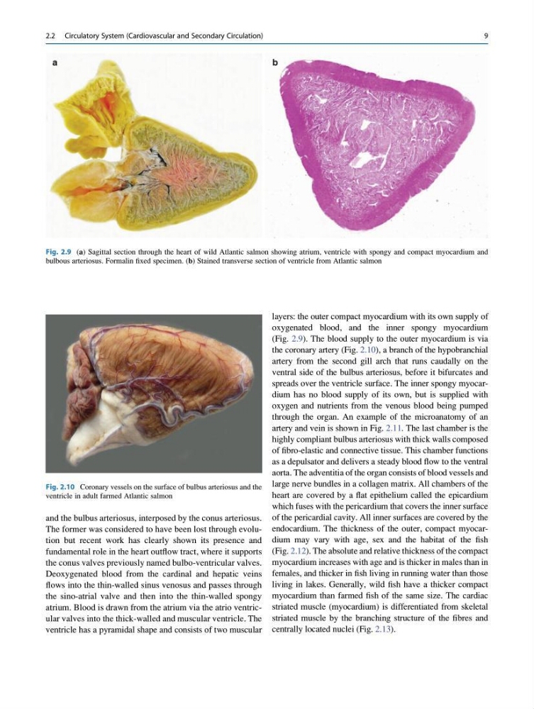
ventricle has a pyramidal shape and consists of two muscular
layers: the outer compact myocardium with its own supply of
oxygenated blood, and the inner spongy myocardium
(Fig. 2.9). The blood supply to the outer myocardium is via
the coronary artery (Fig. 2.10), a branch of the hypobranchial
artery from the second gill arch that runs caudally on the
ventral side of the bulbus arteriosus, before it bifurcates and
spreads over the ventricle surface. The inner spongy myocardium has no blood supply of its own, but is supplied with
oxygen and nutrients from the venous blood being pumped
through the organ. An example of the microanatomy of an
artery and vein is shown in Fig. 2.11. The last chamber is the
highly compliant bulbus arteriosus with thick walls composed
of fibro-elastic and connective tissue. This chamber functions
as a depulsator and delivers a steady blood flow to the ventral
aorta. The adventitia of the organ consists of blood vessels and
large nerve bundles in a collagen matrix. All chambers of the
heart are covered by a flat epithelium called the epicardium
which fuses with the pericardium that covers the inner surface
of the pericardial cavity. All inner surfaces are covered by the
endocardium. The thickness of the outer, compact myocardium may vary with age, sex and the habitat of the fish
(Fig. 2.12). The absolute and relative thickness of the compact
myocardium increases with age and is thicker in males than in
females, and thicker in fish living in running water than those
living in lakes. Generally, wild fish have a thicker compact
myocardium than farmed fish of the same size. The cardiac
striated muscle (myocardium) is differentiated from skeletal
striated muscle by the branching structure of the fibres and
centrally located nuclei (Fig. 2.13).
Fig. 2.9 (a) Sagittal section through the heart of wild Atlantic salmon showing atrium, ventricle with spongy and compact myocardium and
bulbous arteriosus. Formalin fixed specimen. (b) Stained transverse section of ventricle from Atlantic salmon
Fig. 2.10 Coronary vessels on the surface of bulbus arteriosus and the
ventricle in adult farmed Atlantic salmon
2.2 Circulatory System (Cardiovascular and Secondary Circulation)
9
The former was considered to have been lost through evolution but recent work has clearly shown its presence and
fundamental role in the heart outflow tract, where it supports
the conus valves previously named bulbo-ventricular valves.
Deoxygenated blood from the cardinal and hepatic veins
flows into the thin-walled sinus venosus and passes through
the sino-atrial valve and then into the thin-walled spongy
atrium. Blood is drawn from the atrium via the atrio ventricular valves into the thick-walled and muscular ventricle. The
ventricle has a pyramidal shape and consists of two muscular
layers: the outer compact myocardium with its own supply of
oxygenated blood, and the inner spongy myocardium
(Fig. 2.9). The blood supply to the outer myocardium is via
the coronary artery (Fig. 2.10), a branch of the hypobranchial
artery from the second gill arch that runs caudally on the
ventral side of the bulbus arteriosus, before it bifurcates and
spreads over the ventricle surface. The inner spongy myocardium has no blood supply of its own, but is supplied with
oxygen and nutrients from the venous blood being pumped
through the organ. An example of the microanatomy of an
artery and vein is shown in Fig. 2.11. The last chamber is the
highly compliant bulbus arteriosus with thick walls composed
of fibro-elastic and connective tissue. This chamber functions
as a depulsator and delivers a steady blood flow to the ventral
aorta. The adventitia of the organ consists of blood vessels and
large nerve bundles in a collagen matrix. All chambers of the
heart are covered by a flat epithelium called the epicardium
which fuses with the pericardium that covers the inner surface
of the pericardial cavity. All inner surfaces are covered by the
endocardium. The thickness of the outer, compact myocardium may vary with age, sex and the habitat of the fish
(Fig. 2.12). The absolute and relative thickness of the compact
myocardium increases with age and is thicker in males than in
females, and thicker in fish living in running water than those
living in lakes. Generally, wild fish have a thicker compact
myocardium than farmed fish of the same size. The cardiac
striated muscle (myocardium) is differentiated from skeletal
striated muscle by the branching structure of the fibres and
centrally located nuclei (Fig. 2.13).
Fig. 2.9 (a) Sagittal section through the heart of wild Atlantic salmon showing atrium, ventricle with spongy and compact myocardium and
bulbous arteriosus. Formalin fixed specimen. (b) Stained transverse section of ventricle from Atlantic salmon
Fig. 2.10 Coronary vessels on the surface of bulbus arteriosus and the
ventricle in adult farmed Atlantic salmon
2.2 Circulatory System (Cardiovascular and Secondary Circulation)
9