

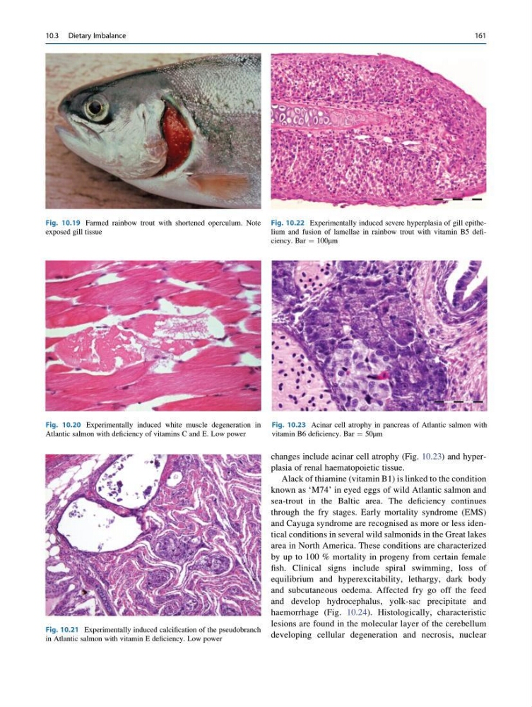
changes include acinar cell atrophy (Fig. 10.23) and hyperplasia of renal haematopoietic tissue.
Alack of thiamine (vitamin B1) is linked to the condition
known as ‘M74’ in eyed eggs of wild Atlantic salmon and
sea-trout in the Baltic area. The deficiency continues
through the fry stages. Early mortality syndrome (EMS)
and Cayuga syndrome are recognised as more or less identical conditions in several wild salmonids in the Great lakes
area in North America. These conditions are characterized
by up to 100 % mortality in progeny from certain female
fish. Clinical signs include spiral swimming, loss of
equilibrium and hyperexcitability, lethargy, dark body
and subcutaneous oedema. Affected fry go off the feed
and develop hydrocephalus, yolk-sac precipitate and
haemorrhage (Fig. 10.24). Histologically, characteristic
lesions are found in the molecular layer of the cerebellum
developing cellular degeneration and necrosis, nuclear
Fig. 10.19 Farmed rainbow trout with shortened operculum. Note
exposed gill tissue
Fig. 10.20 Experimentally induced white muscle degeneration in
Atlantic salmon with deficiency of vitamins C and E. Low power
Fig. 10.21 Experimentally induced calcification of the pseudobranch
in Atlantic salmon with vitamin E deficiency. Low power
Fig. 10.22 Experimentally induced severe hyperplasia of gill epithelium and fusion of lamellae in rainbow trout with vitamin B5 deficiency. Bar ¼ 100μm
Fig. 10.23 Acinar cell atrophy in pancreas of Atlantic salmon with
vitamin B6 deficiency. Bar ¼ 50μm
10.3 Dietary Imbalance
161
Alack of thiamine (vitamin B1) is linked to the condition
known as ‘M74’ in eyed eggs of wild Atlantic salmon and
sea-trout in the Baltic area. The deficiency continues
through the fry stages. Early mortality syndrome (EMS)
and Cayuga syndrome are recognised as more or less identical conditions in several wild salmonids in the Great lakes
area in North America. These conditions are characterized
by up to 100 % mortality in progeny from certain female
fish. Clinical signs include spiral swimming, loss of
equilibrium and hyperexcitability, lethargy, dark body
and subcutaneous oedema. Affected fry go off the feed
and develop hydrocephalus, yolk-sac precipitate and
haemorrhage (Fig. 10.24). Histologically, characteristic
lesions are found in the molecular layer of the cerebellum
developing cellular degeneration and necrosis, nuclear
Fig. 10.19 Farmed rainbow trout with shortened operculum. Note
exposed gill tissue
Fig. 10.20 Experimentally induced white muscle degeneration in
Atlantic salmon with deficiency of vitamins C and E. Low power
Fig. 10.21 Experimentally induced calcification of the pseudobranch
in Atlantic salmon with vitamin E deficiency. Low power
Fig. 10.22 Experimentally induced severe hyperplasia of gill epithelium and fusion of lamellae in rainbow trout with vitamin B5 deficiency. Bar ¼ 100μm
Fig. 10.23 Acinar cell atrophy in pancreas of Atlantic salmon with
vitamin B6 deficiency. Bar ¼ 50μm
10.3 Dietary Imbalance
161