

8.4.2 Loma salmonae
Loma salmonae is an economical important obligate gill
pathogen. It has been detected from farmed rainbow trout,
Chinook, coho and sockeye salmon reared in fresh water,
and among some species reared in sea water cages. Affected
fish may show respiratory distress and impaired swimming,
with reduced growth rates. Gross signs include exophthalmia, ascites and petechiae on the opercula. Small,
round, white cyst-like formations (xenoma) up to 0.5 mm
in diameter can be observed within various tissues, but
principally in the gill lamellae and generally in close association with the pillar cell system, during sporogony. The
target cells for L. salmonae include pillar and endothelial
cells, or leucocytes that migrate through the basement membrane of a blood vessel. Significant pathological changes
occur as the infected cell undergoes hypertrophy and marked
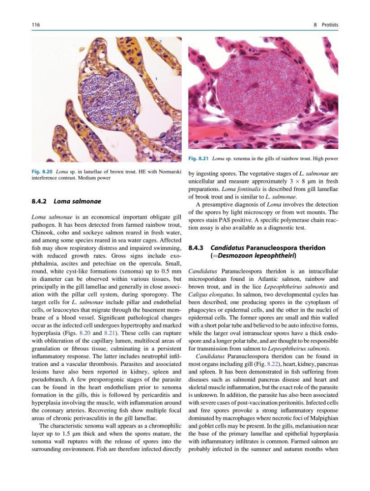
hyperplasia (Figs. 8.20 and 8.21). These cells can rupture
with obliteration of the capillary lumen, multifocal areas of
granulation or fibrous tissue, culminating in a persistent
inflammatory response. The latter includes neutrophil infiltration and a vascular thrombosis. Parasites and associated
lesions have also been reported in kidney, spleen and
pseudobranch. A few presporogonic stages of the parasite
can be found in the heart endothelium prior to xenoma
formation in the gills, this is followed by pericarditis and
hyperplasia involving the muscle, with inflammation around
the coronary arteries. Recovering fish show multiple focal
areas of chronic perivasculitis in the gill lamellae.
The characteristic xenoma wall appears as a chromophilic
layer up to 1.5 μm thick and when the spores mature, the
xenoma wall ruptures with the release of spores into the
surrounding environment. Fish are therefore infected directly
by ingesting spores. The vegetative stages of L. salmonae are
unicellular and measure approximately 3 Â 8 μm in fresh
preparations. Loma fontinalis is described from gill lamellae
of brook trout and is similar to L. salmonae.
A presumptive diagnosis of Loma involves the detection
of the spores by light microscopy or from wet mounts. The
spores stain PAS positive. A specific polymerase chain reaction assay is also available as a diagnostic test.
8.4.3 Candidatus Paranucleospora theridon
(¼Desmozoon lepeophtheiri)
Candidatus Paranucleospora theridon is an intracellular
microsporidean found in Atlantic salmon, rainbow and
brown trout, and in the lice Lepeophtheirus salmonis and
Caligus elongatus. In salmon, two developmental cycles has
been described, one producing spores in the cytoplasm of
phagocytes or epidermal cells, and the other in the nuclei of
epidermal cells. The former spores are small and thin walled
with a short polar tube and believed to be auto infective forms,
while the larger oval intranuclear spores have a thick endospore and a longer polar tube, and are thought to be responsible
for transmission from salmon to Lepeophtheirus salmonis.
Candidatus Paranucleospora theridon can be found in
most organs including gill (Fig. 8.22), heart, kidney, pancreas
and spleen. It has been demonstrated in fish suffering from
diseases such as salmonid pancreas disease and heart and
skeletal muscle inflammation, but the exact role of the parasite
is unknown. In addition, the parasite has also been associated
with severe cases of post-vaccination peritonitis. Infected cells
and free spores provoke a strong inflammatory response
dominated by macrophages where necrotic foci of Malpighian
and goblet cells may be present. In the gills, melanisation near
the base of the primary lamellae and epithelial hyperplasia
with inflammatory infiltrates is common. Farmed salmon are
probably infected in the summer and autumn months when
Fig. 8.20 Loma sp. in lamellae of brown trout. HE with Normarski
interference contrast. Medium power
Fig. 8.21 Loma sp. xenoma in the gills of rainbow trout. High power
116
8 Protists
Loma salmonae is an economical important obligate gill
pathogen. It has been detected from farmed rainbow trout,
Chinook, coho and sockeye salmon reared in fresh water,
and among some species reared in sea water cages. Affected
fish may show respiratory distress and impaired swimming,
with reduced growth rates. Gross signs include exophthalmia, ascites and petechiae on the opercula. Small,
round, white cyst-like formations (xenoma) up to 0.5 mm
in diameter can be observed within various tissues, but
principally in the gill lamellae and generally in close association with the pillar cell system, during sporogony. The
target cells for L. salmonae include pillar and endothelial
cells, or leucocytes that migrate through the basement membrane of a blood vessel. Significant pathological changes
occur as the infected cell undergoes hypertrophy and marked
hyperplasia (Figs. 8.20 and 8.21). These cells can rupture
with obliteration of the capillary lumen, multifocal areas of
granulation or fibrous tissue, culminating in a persistent
inflammatory response. The latter includes neutrophil infiltration and a vascular thrombosis. Parasites and associated
lesions have also been reported in kidney, spleen and
pseudobranch. A few presporogonic stages of the parasite
can be found in the heart endothelium prior to xenoma
formation in the gills, this is followed by pericarditis and
hyperplasia involving the muscle, with inflammation around
the coronary arteries. Recovering fish show multiple focal
areas of chronic perivasculitis in the gill lamellae.
The characteristic xenoma wall appears as a chromophilic
layer up to 1.5 μm thick and when the spores mature, the
xenoma wall ruptures with the release of spores into the
surrounding environment. Fish are therefore infected directly
by ingesting spores. The vegetative stages of L. salmonae are
unicellular and measure approximately 3 Â 8 μm in fresh
preparations. Loma fontinalis is described from gill lamellae
of brook trout and is similar to L. salmonae.
A presumptive diagnosis of Loma involves the detection
of the spores by light microscopy or from wet mounts. The
spores stain PAS positive. A specific polymerase chain reaction assay is also available as a diagnostic test.
8.4.3 Candidatus Paranucleospora theridon
(¼Desmozoon lepeophtheiri)
Candidatus Paranucleospora theridon is an intracellular
microsporidean found in Atlantic salmon, rainbow and
brown trout, and in the lice Lepeophtheirus salmonis and
Caligus elongatus. In salmon, two developmental cycles has
been described, one producing spores in the cytoplasm of
phagocytes or epidermal cells, and the other in the nuclei of
epidermal cells. The former spores are small and thin walled
with a short polar tube and believed to be auto infective forms,
while the larger oval intranuclear spores have a thick endospore and a longer polar tube, and are thought to be responsible
for transmission from salmon to Lepeophtheirus salmonis.
Candidatus Paranucleospora theridon can be found in
most organs including gill (Fig. 8.22), heart, kidney, pancreas
and spleen. It has been demonstrated in fish suffering from
diseases such as salmonid pancreas disease and heart and
skeletal muscle inflammation, but the exact role of the parasite
is unknown. In addition, the parasite has also been associated
with severe cases of post-vaccination peritonitis. Infected cells
and free spores provoke a strong inflammatory response
dominated by macrophages where necrotic foci of Malpighian
and goblet cells may be present. In the gills, melanisation near
the base of the primary lamellae and epithelial hyperplasia
with inflammatory infiltrates is common. Farmed salmon are
probably infected in the summer and autumn months when
Fig. 8.20 Loma sp. in lamellae of brown trout. HE with Normarski
interference contrast. Medium power
Fig. 8.21 Loma sp. xenoma in the gills of rainbow trout. High power
116
8 Protists