

8.4
Microsporidia
8.4.1 Kabatana takedai
Kabatana takedai (formally Microsporidium takedi) affects
the muscle and heart of various species including chum,
masou, pink and sockeye salmon, rainbow and brown trout,
and Japanese char. A seasonal prevalence of this parasite is
recognised, with the initial outbreak during the summer at
around 15
C.
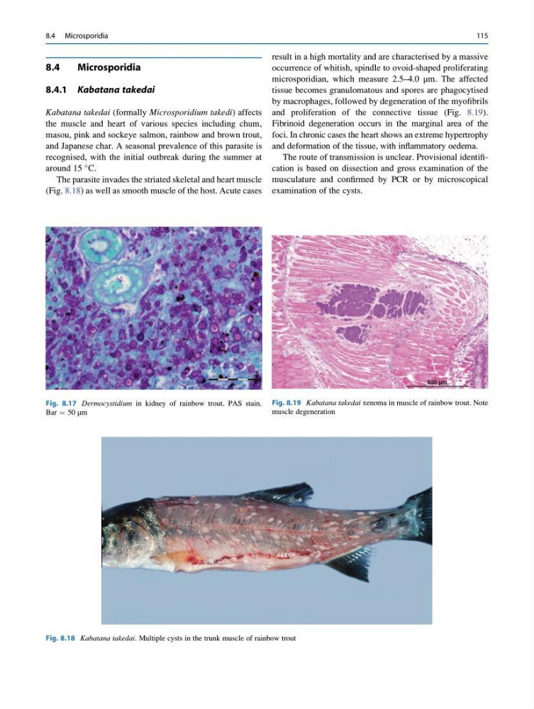
The parasite invades the striated skeletal and heart muscle
(Fig. 8.18) as well as smooth muscle of the host. Acute cases
result in a high mortality and are characterised by a massive
occurrence of whitish, spindle to ovoid-shaped proliferating
microsporidian, which measure 2.5–4.0 μm. The affected
tissue becomes granulomatous and spores are phagocytised
by macrophages, followed by degeneration of the myofibrils
and proliferation of the connective tissue (Fig. 8.19).
Fibrinoid degeneration occurs in the marginal area of the
foci. In chronic cases the heart shows an extreme hypertrophy
and deformation of the tissue, with inflammatory oedema.
The route of transmission is unclear. Provisional identification is based on dissection and gross examination of the
musculature and confirmed by PCR or by microscopical
examination of the cysts.
Fig. 8.17 Dermocystidium in kidney of rainbow trout. PAS stain.
Bar ¼ 50 μm
Fig. 8.18 Kabatana takedai. Multiple cysts in the trunk muscle of rainbow trout
Fig. 8.19 Kabatana takedai xenoma in muscle of rainbow trout. Note
muscle degeneration
8.4 Microsporidia
115
Microsporidia
8.4.1 Kabatana takedai
Kabatana takedai (formally Microsporidium takedi) affects
the muscle and heart of various species including chum,
masou, pink and sockeye salmon, rainbow and brown trout,
and Japanese char. A seasonal prevalence of this parasite is
recognised, with the initial outbreak during the summer at
around 15
C.
The parasite invades the striated skeletal and heart muscle
(Fig. 8.18) as well as smooth muscle of the host. Acute cases
result in a high mortality and are characterised by a massive
occurrence of whitish, spindle to ovoid-shaped proliferating
microsporidian, which measure 2.5–4.0 μm. The affected
tissue becomes granulomatous and spores are phagocytised
by macrophages, followed by degeneration of the myofibrils
and proliferation of the connective tissue (Fig. 8.19).
Fibrinoid degeneration occurs in the marginal area of the
foci. In chronic cases the heart shows an extreme hypertrophy
and deformation of the tissue, with inflammatory oedema.
The route of transmission is unclear. Provisional identification is based on dissection and gross examination of the
musculature and confirmed by PCR or by microscopical
examination of the cysts.
Fig. 8.17 Dermocystidium in kidney of rainbow trout. PAS stain.
Bar ¼ 50 μm
Fig. 8.18 Kabatana takedai. Multiple cysts in the trunk muscle of rainbow trout
Fig. 8.19 Kabatana takedai xenoma in muscle of rainbow trout. Note
muscle degeneration
8.4 Microsporidia
115