

parasite is variable but generally, it is sac-like (50–100 μm),
elongated structure with 10–35 tentacles projecting from the
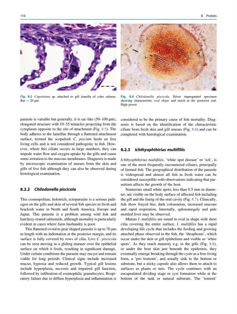
cytoplasm opposite to the site of attachment (Fig. 8.5). The
body adheres to the lamellae through a flattened attachment
surface, termed the scopuloid. C. piscium feeds on free
living cells and is not considered pathogenic to fish. However, where this ciliate occurs in large numbers, they can
impede water flow and oxygen uptake by the gills and cause
some irritation to the mucous membranes. Diagnosis is made
by microscopic examination of smears from the skin and
gills of live fish although they can also be observed during
histological examination.
8.2.2 Chilodonella piscicola
This cosmopolitan, holotrich, ectoparasite is a serious pathogen on the gills and skin of several fish species in fresh and
brackish water in North and South America, Europe and
Japan. This parasite is a problem among wild fish and
hatchery-reared salmonids, although mortality is particularly
evident in cases where farm husbandry is poor.
This flattened ovoid to pear shaped parasite is up to 70 μm
in length with an indentation at the posterior margin, and its
surface is fully covered by rows of cilia. Live C. piscicola
can be seen moving in a gliding manner over the epithelial
surface on which it feeds, resulting in significant damage.
Under certain conditions the parasite may encyst and remain
viable for long periods. Clinical signs include increased
mucus, hypoxia and reduced growth. Typical gill lesions
include hyperplasia, necrosis and impaired gill function,
followed by infiltration of eosinophilic granulocytes. Respiratory failure due to diffuse hyperplasia and inflammation is
considered to be the primary cause of fish mortality. Diagnosis is based on the identification of the characteristic
ciliate from fresh skin and gill smears (Fig. 8.6) and can be
completed with histological examination.
8.2.3 Ichthyophthirius multifiliis
Ichthyophthirius multifiliis, ‘white spot disease’ or ‘ich’, is
one of the most frequently encountered ciliates, principally
of farmed fish. The geographical distribution of the parasite
is widespread and almost all fish in fresh water can be
considered susceptible with observations indicating that parasitism affects the growth of the host.
Numerous small white spots, less than 0.5 mm in diameter, are visible on the body surface of affected fish including
the gill and the lining of the oral cavity (Fig. 8.7). Clinically,
fish show frayed fins, dark colouration, increased mucous
and rapid respiration. Internally, splenomegaly and pale
mottled liver may be observed.
Mature I. multifiliis are round to oval in shape with short
cilia covering the entire surface. I. multifiliis has a rapid
developing life cycle that includes the feeding and growing
attached phase observed in the fish, the ‘throphonts’, which
occur under the skin or gill epithelium and visible as ‘white
spots’. As they reach maturity e.g. in the gills (Fig. 8.8),
or under the host skin just beneath the epidermis, they
eventually emerge breaking through the cysts as a free-living
form, a ‘pro tromont’, and usually sink to the bottom or
substrate, but a sticky capsule also allows them to attach to
surfaces as plants or nets. The cycle continues with an
encapsulated dividing stage or cyst formation while at the
bottom of the tank or natural substrate. The ‘tomont’
Fig. 8.5 Capriniana sp. attached to gill lamella of coho salmon.
Bar ¼ 20 μm
Fig. 8.6 Chilodonella piscicola. Silver impregnated specimen
showing characteristic oval shape and notch at the posterior end.
High power
110
8 Protists
elongated structure with 10–35 tentacles projecting from the
cytoplasm opposite to the site of attachment (Fig. 8.5). The
body adheres to the lamellae through a flattened attachment
surface, termed the scopuloid. C. piscium feeds on free
living cells and is not considered pathogenic to fish. However, where this ciliate occurs in large numbers, they can
impede water flow and oxygen uptake by the gills and cause
some irritation to the mucous membranes. Diagnosis is made
by microscopic examination of smears from the skin and
gills of live fish although they can also be observed during
histological examination.
8.2.2 Chilodonella piscicola
This cosmopolitan, holotrich, ectoparasite is a serious pathogen on the gills and skin of several fish species in fresh and
brackish water in North and South America, Europe and
Japan. This parasite is a problem among wild fish and
hatchery-reared salmonids, although mortality is particularly
evident in cases where farm husbandry is poor.
This flattened ovoid to pear shaped parasite is up to 70 μm
in length with an indentation at the posterior margin, and its
surface is fully covered by rows of cilia. Live C. piscicola
can be seen moving in a gliding manner over the epithelial
surface on which it feeds, resulting in significant damage.
Under certain conditions the parasite may encyst and remain
viable for long periods. Clinical signs include increased
mucus, hypoxia and reduced growth. Typical gill lesions
include hyperplasia, necrosis and impaired gill function,
followed by infiltration of eosinophilic granulocytes. Respiratory failure due to diffuse hyperplasia and inflammation is
considered to be the primary cause of fish mortality. Diagnosis is based on the identification of the characteristic
ciliate from fresh skin and gill smears (Fig. 8.6) and can be
completed with histological examination.
8.2.3 Ichthyophthirius multifiliis
Ichthyophthirius multifiliis, ‘white spot disease’ or ‘ich’, is
one of the most frequently encountered ciliates, principally
of farmed fish. The geographical distribution of the parasite
is widespread and almost all fish in fresh water can be
considered susceptible with observations indicating that parasitism affects the growth of the host.
Numerous small white spots, less than 0.5 mm in diameter, are visible on the body surface of affected fish including
the gill and the lining of the oral cavity (Fig. 8.7). Clinically,
fish show frayed fins, dark colouration, increased mucous
and rapid respiration. Internally, splenomegaly and pale
mottled liver may be observed.
Mature I. multifiliis are round to oval in shape with short
cilia covering the entire surface. I. multifiliis has a rapid
developing life cycle that includes the feeding and growing
attached phase observed in the fish, the ‘throphonts’, which
occur under the skin or gill epithelium and visible as ‘white
spots’. As they reach maturity e.g. in the gills (Fig. 8.8),
or under the host skin just beneath the epidermis, they
eventually emerge breaking through the cysts as a free-living
form, a ‘pro tromont’, and usually sink to the bottom or
substrate, but a sticky capsule also allows them to attach to
surfaces as plants or nets. The cycle continues with an
encapsulated dividing stage or cyst formation while at the
bottom of the tank or natural substrate. The ‘tomont’
Fig. 8.5 Capriniana sp. attached to gill lamella of coho salmon.
Bar ¼ 20 μm
Fig. 8.6 Chilodonella piscicola. Silver impregnated specimen
showing characteristic oval shape and notch at the posterior end.
High power
110
8 Protists