

within membrane-bound cytoplasmic vacuoles using H&E,
methylene blue or Giemsa stained sections, and provide a
presumptive diagnosis. Similarly, macrophages containing
P. salmonis can be detected in stained peripheral blood
imprints. P. salmonis is a Gram negative, acid-fast, nonmotile, predominantly coccoid, non-capsulated (although
often pleomorphic) organism. Several cell lines including
CHSE-214 and RTG-2 and antibiotic free media, have been
used successfully for the primary isolation of P. salmonis from
kidney. Confirmation can be achieved through the use of an
ELISA, presence in cell culture, a PCR assay or in situ
hybridization. Culture media including a marine based broth
supplemented with L-cysteine appears to allow the successful
culture independently of the more costly and time consuming
isolation on cell lines. Additionally, it avoids the difficulty of
eliminating the contamination with host cell debris. A differential diagnosis would include viral haemorrhagic septicaemia.
6.21 Epitheliocystis
At least three bacterial species may be involved in the
condition known as epitheliocystis, two of which,
Candidatus Piscichlamydia salmonis which is mainly
from sea waterfish in Ireland and Norway, and Candidatus
Clavochlamydia salmonicola from freshwater fish, belong to
the Phylum Chlamydiae. This group is a cosmopolitan class
of intracellular granular, basophilic Gram-negative bacteria,
considered mostly as opportunistic rather than primary
pathogens. Both species have been reported from wild freshwater brown trout in Switzerland, and Candidatus
Piscichlamydia salmonis has been diagnosed in freshwaterreared Arctic char in North America and in-farmed Atlantic
salmon in Ireland and Norway. Clavochlamydia salmonicola
related epitheliocysts have been reported to disappear
6 weeks after transfer to sea water. As gill diseases often
have a complex aetiology, the exact role of the different
pathogens and environmental factors involved may be difficult to ascertain.
Clinical signs in affected fish include lethargy induced by
the severe hyperplastic gill inflammation, leading to hyperventilation, flared opercula and increased mucus production.
Mortality levels in sea-farmed salmon may be highly variable but up to 80 % has been recorded, although this also
depends on environmental conditions and the concurrence of
other pathogens. A seasonal occurrence typically peaking in
the autumn months indicate water temperature as an important risk factor.
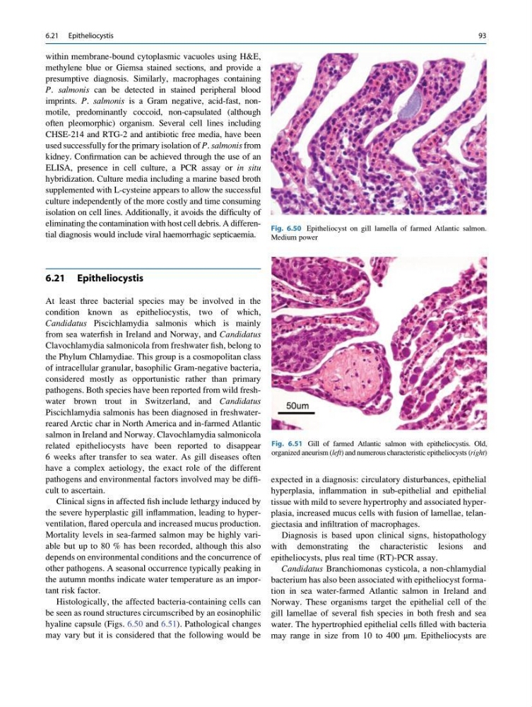
Histologically, the affected bacteria-containing cells can
be seen as round structures circumscribed by an eosinophilic
hyaline capsule (Figs. 6.50 and 6.51). Pathological changes
may vary but it is considered that the following would be
expected in a diagnosis: circulatory disturbances, epithelial
hyperplasia, inflammation in sub-epithelial and epithelial
tissue with mild to severe hypertrophy and associated hyperplasia, increased mucus cells with fusion of lamellae, telangiectasia and infiltration of macrophages.
Diagnosis is based upon clinical signs, histopathology
with demonstrating the characteristic lesions and
epitheliocysts, plus real time (RT)-PCR assay.
Candidatus Branchiomonas cysticola, a non-chlamydial
bacterium has also been associated with epitheliocyst formation in sea water-farmed Atlantic salmon in Ireland and
Norway. These organisms target the epithelial cell of the
gill lamellae of several fish species in both fresh and sea
water. The hypertrophied epithelial cells filled with bacteria
may range in size from 10 to 400 μm. Epitheliocysts are
Fig. 6.51 Gill of farmed Atlantic salmon with epitheliocystis. Old,
organized aneurism (left) and numerous characteristic epitheliocysts (right)
Fig. 6.50 Epitheliocyst on gill lamella of farmed Atlantic salmon.
Medium power
6.21 Epitheliocystis
93
methylene blue or Giemsa stained sections, and provide a
presumptive diagnosis. Similarly, macrophages containing
P. salmonis can be detected in stained peripheral blood
imprints. P. salmonis is a Gram negative, acid-fast, nonmotile, predominantly coccoid, non-capsulated (although
often pleomorphic) organism. Several cell lines including
CHSE-214 and RTG-2 and antibiotic free media, have been
used successfully for the primary isolation of P. salmonis from
kidney. Confirmation can be achieved through the use of an
ELISA, presence in cell culture, a PCR assay or in situ
hybridization. Culture media including a marine based broth
supplemented with L-cysteine appears to allow the successful
culture independently of the more costly and time consuming
isolation on cell lines. Additionally, it avoids the difficulty of
eliminating the contamination with host cell debris. A differential diagnosis would include viral haemorrhagic septicaemia.
6.21 Epitheliocystis
At least three bacterial species may be involved in the
condition known as epitheliocystis, two of which,
Candidatus Piscichlamydia salmonis which is mainly
from sea waterfish in Ireland and Norway, and Candidatus
Clavochlamydia salmonicola from freshwater fish, belong to
the Phylum Chlamydiae. This group is a cosmopolitan class
of intracellular granular, basophilic Gram-negative bacteria,
considered mostly as opportunistic rather than primary
pathogens. Both species have been reported from wild freshwater brown trout in Switzerland, and Candidatus
Piscichlamydia salmonis has been diagnosed in freshwaterreared Arctic char in North America and in-farmed Atlantic
salmon in Ireland and Norway. Clavochlamydia salmonicola
related epitheliocysts have been reported to disappear
6 weeks after transfer to sea water. As gill diseases often
have a complex aetiology, the exact role of the different
pathogens and environmental factors involved may be difficult to ascertain.
Clinical signs in affected fish include lethargy induced by
the severe hyperplastic gill inflammation, leading to hyperventilation, flared opercula and increased mucus production.
Mortality levels in sea-farmed salmon may be highly variable but up to 80 % has been recorded, although this also
depends on environmental conditions and the concurrence of
other pathogens. A seasonal occurrence typically peaking in
the autumn months indicate water temperature as an important risk factor.
Histologically, the affected bacteria-containing cells can
be seen as round structures circumscribed by an eosinophilic
hyaline capsule (Figs. 6.50 and 6.51). Pathological changes
may vary but it is considered that the following would be
expected in a diagnosis: circulatory disturbances, epithelial
hyperplasia, inflammation in sub-epithelial and epithelial
tissue with mild to severe hypertrophy and associated hyperplasia, increased mucus cells with fusion of lamellae, telangiectasia and infiltration of macrophages.
Diagnosis is based upon clinical signs, histopathology
with demonstrating the characteristic lesions and
epitheliocysts, plus real time (RT)-PCR assay.
Candidatus Branchiomonas cysticola, a non-chlamydial
bacterium has also been associated with epitheliocyst formation in sea water-farmed Atlantic salmon in Ireland and
Norway. These organisms target the epithelial cell of the
gill lamellae of several fish species in both fresh and sea
water. The hypertrophied epithelial cells filled with bacteria
may range in size from 10 to 400 μm. Epitheliocysts are
Fig. 6.51 Gill of farmed Atlantic salmon with epitheliocystis. Old,
organized aneurism (left) and numerous characteristic epitheliocysts (right)
Fig. 6.50 Epitheliocyst on gill lamella of farmed Atlantic salmon.
Medium power
6.21 Epitheliocystis
93