

36 Microarrays and DNA-Chips in the Analysis of Genetic Aberrations
481
Analysis of DNA extracted from the breast cancer cell line BT474
(ATCC No. HTB-20) with our cDNA arrays shows the known amplification of ERBB2 (6 fold) (Pinkel et al. 1998, Pollack et al. 1999) in Figure 3.
Our fmding of a 6 fold amplification for the gene CYP24 confirms a recent
report (Albertson et al. 2000) that identifies CYP24 gene as a candidate
oncogene located on an approximately 2-Mb amplified (8-12X) region
on chromosome 20q13.2. Note that this cell line has an average number
of 103 chromosomes/cell, meaning that because of our normalization algorythm copy numbers for "amplified" genes are likely to be underestimated.
Troubleshooting
As for any in situ hybridization protocol, failure ofthe assay translates into
poor or no signal on all or part of the target spots. One of the major problem areas is the extraction and labeling of sample DNA, which can not be
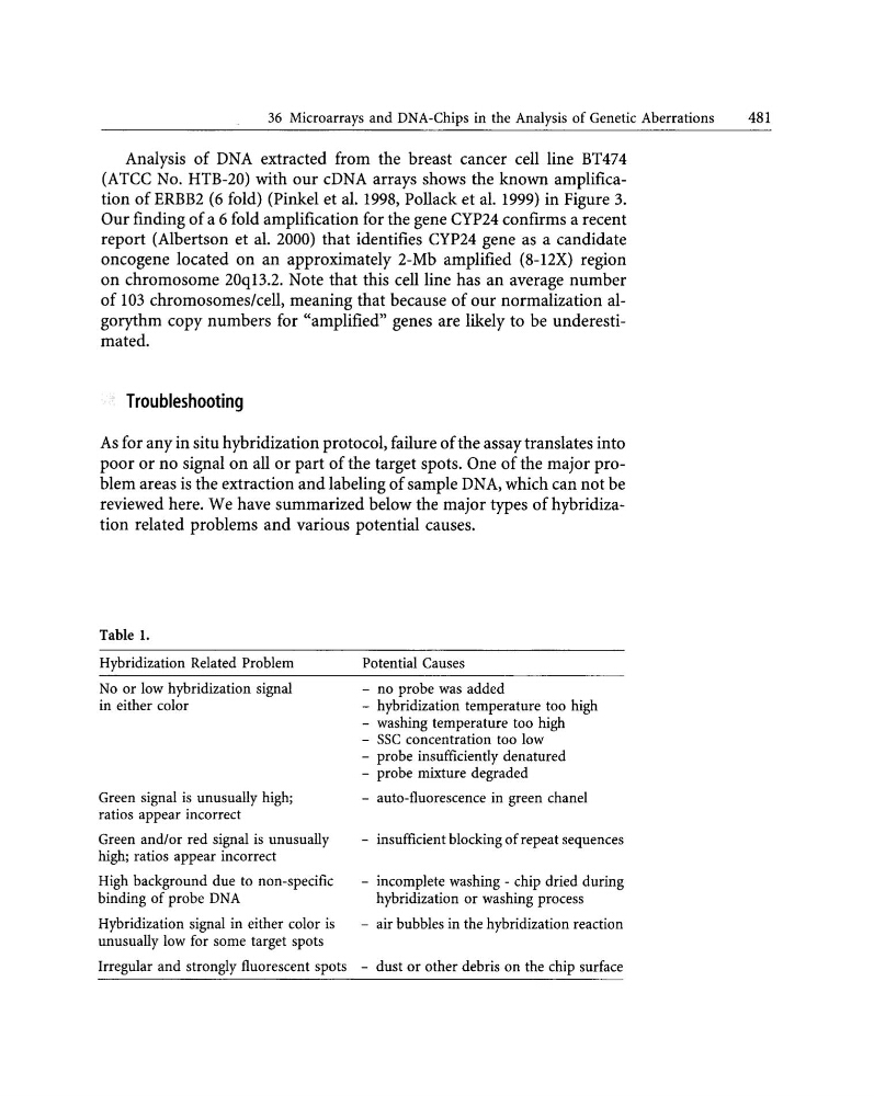
reviewed here. We have summarized below the major types of hybridization related problems and various potential causes.
Table 1.
Hybridization Related Problem
No or low hybridization signal
in either color
Green signal is unusually high;
ratios appear incorrect
Green and/or red signal is unusually
high; ratios appear incorrect
High background due to non-specific
binding of probe DNA
Hybridization signal in either color is
unusually low for some target spots
Irregular and strongly fluorescent spots
Potential Causes
- no probe was added
- hybridization temperature too high
- washing temperature too high
- SSC concentration too low
- probe insufficiently denatured
- probe mixture degraded
- auto-fluorescence in green chanel
- insufficient blocking of repeat sequences
- incomplete washing - chip dried during
hybridization or washing process
- air bubbles in the hybridization reaction
- dust or other debris on the chip surface
481
Analysis of DNA extracted from the breast cancer cell line BT474
(ATCC No. HTB-20) with our cDNA arrays shows the known amplification of ERBB2 (6 fold) (Pinkel et al. 1998, Pollack et al. 1999) in Figure 3.
Our fmding of a 6 fold amplification for the gene CYP24 confirms a recent
report (Albertson et al. 2000) that identifies CYP24 gene as a candidate
oncogene located on an approximately 2-Mb amplified (8-12X) region
on chromosome 20q13.2. Note that this cell line has an average number
of 103 chromosomes/cell, meaning that because of our normalization algorythm copy numbers for "amplified" genes are likely to be underestimated.
Troubleshooting
As for any in situ hybridization protocol, failure ofthe assay translates into
poor or no signal on all or part of the target spots. One of the major problem areas is the extraction and labeling of sample DNA, which can not be
reviewed here. We have summarized below the major types of hybridization related problems and various potential causes.
Table 1.
Hybridization Related Problem
No or low hybridization signal
in either color
Green signal is unusually high;
ratios appear incorrect
Green and/or red signal is unusually
high; ratios appear incorrect
High background due to non-specific
binding of probe DNA
Hybridization signal in either color is
unusually low for some target spots
Irregular and strongly fluorescent spots
Potential Causes
- no probe was added
- hybridization temperature too high
- washing temperature too high
- SSC concentration too low
- probe insufficiently denatured
- probe mixture degraded
- auto-fluorescence in green chanel
- insufficient blocking of repeat sequences
- incomplete washing - chip dried during
hybridization or washing process
- air bubbles in the hybridization reaction
- dust or other debris on the chip surface