

36 Microarrays and DNA-Chips in the Analysis of Genetic Aberrations
479
Results
The Coming cancer array contains approximately 1700 unique cDNAs in 2
replicates and is designed for measuring gene expression profiles and
genomic amplifications in tumor DNA. We have tested the performance
of these arrays in CGH applications using two well-studied tumor cell
lines. Our hybridizations were carried out with a mixture of total human
reference DNA (labeled with Cy3) and DNA extracted from the tumor cell
line (labeled with Cy5).
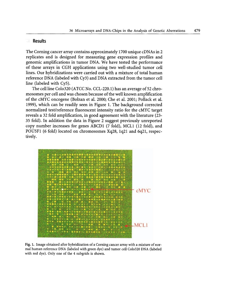
The cell line Colo320 (ATCC No. CCL-220.1) has an average of 52 chromosomes per cell and was chosen because of the well known amplification
of the cMYC oncogene (Bolzan et al. 2000; Che et al. 2001; Pollack et al.
1999), which can be readily seen in Figure 1. The background corrected
normalized test/reference fluorescent intensity ratio for the cMYC target
reveals a 32 fold amplification, in good agreement with the literature (2335 fold). In addition the data in Figure 2 suggest previously unreported
copy number increases for genes ABCDI (7 fold), MCLl (12 fold), and
POU5FI (6 fold) located on chromosomes Xq28, Iq21 and 6q2I, respectively.
Ci YC
Fig. 1. Image obtained after hybridization of a Corning cancer array with a mixture of normal human reference DNA (labeled with green dye) and tumor cell Colo320 DNA (labeled
with red dye). Only one of the 4 subgrids is shown.
479
Results
The Coming cancer array contains approximately 1700 unique cDNAs in 2
replicates and is designed for measuring gene expression profiles and
genomic amplifications in tumor DNA. We have tested the performance
of these arrays in CGH applications using two well-studied tumor cell
lines. Our hybridizations were carried out with a mixture of total human
reference DNA (labeled with Cy3) and DNA extracted from the tumor cell
line (labeled with Cy5).
The cell line Colo320 (ATCC No. CCL-220.1) has an average of 52 chromosomes per cell and was chosen because of the well known amplification
of the cMYC oncogene (Bolzan et al. 2000; Che et al. 2001; Pollack et al.
1999), which can be readily seen in Figure 1. The background corrected
normalized test/reference fluorescent intensity ratio for the cMYC target
reveals a 32 fold amplification, in good agreement with the literature (2335 fold). In addition the data in Figure 2 suggest previously unreported
copy number increases for genes ABCDI (7 fold), MCLl (12 fold), and
POU5FI (6 fold) located on chromosomes Xq28, Iq21 and 6q2I, respectively.
Ci YC
Fig. 1. Image obtained after hybridization of a Corning cancer array with a mixture of normal human reference DNA (labeled with green dye) and tumor cell Colo320 DNA (labeled
with red dye). Only one of the 4 subgrids is shown.