

22 DNA Breakage Detection-FISH (DBD-FISH)
289
•
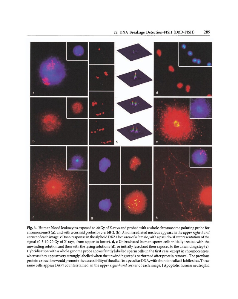
Fig. 3. Human blood leukocytes exposed to 20 Gy of X-rays and probed with a whole chromosome painting probe for
chromosome 8 (a), and with a cosmid probe for c-erbB-2. (b). An unirradiated nucleus appears in the upper right-hand
corner ofeach image. c Dose-response in the alphoid DXZlioci area ofa female, with a pseudo-3D representation ofthe
signal (0-5-10-20 Gy of X-rays, from upper to lower). d, e Unirradiated human sperm cells initially treated with the
unwinding solution and then with the lysing solutions (d), or initially lysed and then exposed to the unwinding step (e).
Hybridisation with a whole genome probe shows faintly labelled sperm cells in the first case, except in chromocentres,
whereas they appear very strongly labelled when the unwinding step is performed after protein removal. The previous
protein extraction would promote the accessibilityofthe alkali to a peculiar DNA, with abundant alkali-labile sites. These
same cells appear DAPI counterstained, in the upper right-hand corner of each image. f Apoptotic human neutrophil
289
•
Fig. 3. Human blood leukocytes exposed to 20 Gy of X-rays and probed with a whole chromosome painting probe for
chromosome 8 (a), and with a cosmid probe for c-erbB-2. (b). An unirradiated nucleus appears in the upper right-hand
corner ofeach image. c Dose-response in the alphoid DXZlioci area ofa female, with a pseudo-3D representation ofthe
signal (0-5-10-20 Gy of X-rays, from upper to lower). d, e Unirradiated human sperm cells initially treated with the
unwinding solution and then with the lysing solutions (d), or initially lysed and then exposed to the unwinding step (e).
Hybridisation with a whole genome probe shows faintly labelled sperm cells in the first case, except in chromocentres,
whereas they appear very strongly labelled when the unwinding step is performed after protein removal. The previous
protein extraction would promote the accessibilityofthe alkali to a peculiar DNA, with abundant alkali-labile sites. These
same cells appear DAPI counterstained, in the upper right-hand corner of each image. f Apoptotic human neutrophil