

11 Formalin-Fixed and Paraffin-Embedded Tissue Sections
157
represents a tissue with a low density of nuclei. In tumors which were
characterized by a high tumor cell density, e.g. lymphomas or ductal breast
carcinomas,S Jlm sections were sufficient for the evaluation of the signal
numbers. The investigations aimed at identifying high level gene amplifications such as the proto-oncogenes HER-2/neu or c-myc in ductal
breast cancer were even hampered by thick sections, so the use of 5 Jlm
sections was preferred.
Troubleshooting
Hybridization efficiency and tissue morphology are usually negatively correlated. Aggressive pretreatment procedures improving hybridization efficiency (e.g. long incubation times of NaSCN or proteolytic digestion)
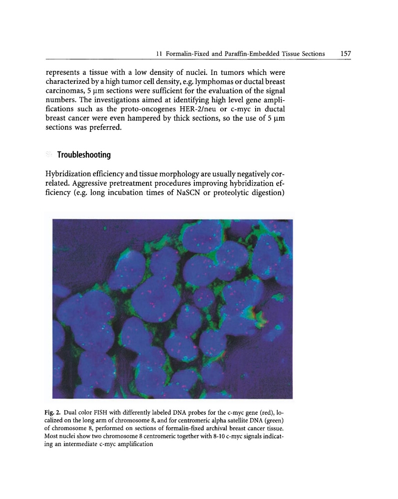
Fig.2. Dual color FISH with differently labeled DNA probes for the c-myc gene (red), localized on the long arm of chromosome 8, and for centromeric alpha satellite DNA (green)
of chromosome 8, performed on sections of formalin-fixed archival breast cancer tissue.
Most nuclei show two chromosome 8 centromeric together with 8-10 c-myc signals indicating an intermediate c-myc amplification
157
represents a tissue with a low density of nuclei. In tumors which were
characterized by a high tumor cell density, e.g. lymphomas or ductal breast
carcinomas,S Jlm sections were sufficient for the evaluation of the signal
numbers. The investigations aimed at identifying high level gene amplifications such as the proto-oncogenes HER-2/neu or c-myc in ductal
breast cancer were even hampered by thick sections, so the use of 5 Jlm
sections was preferred.
Troubleshooting
Hybridization efficiency and tissue morphology are usually negatively correlated. Aggressive pretreatment procedures improving hybridization efficiency (e.g. long incubation times of NaSCN or proteolytic digestion)
Fig.2. Dual color FISH with differently labeled DNA probes for the c-myc gene (red), localized on the long arm of chromosome 8, and for centromeric alpha satellite DNA (green)
of chromosome 8, performed on sections of formalin-fixed archival breast cancer tissue.
Most nuclei show two chromosome 8 centromeric together with 8-10 c-myc signals indicating an intermediate c-myc amplification