

156
HARALD RIEDER et al.
5% of all nuclei corresponded well with the true copy number of the respective chromosome and was independent of section thickness and also
of chromosome count. The fraction of the nuclei which represented the
true chromosome number decreased with the copy number of this chromosome and increased with the section thickness. In placental tissues, for
instance, it was highest in monosomy and lowest in tetrasomy at a section
thickness of 20 /lm. Except for monosomies, the detection of low grade
mosaicism usually required sections thicker than 5 /lm and the evaluation
of an increased number of cells.
The average number of spots per nucleus was also used to identify
monosomies, disomies and trisomies in thin tissue sections, while detection of tetrasomies required sections of more than 15 /lm. The influence of
section thickness on signal distribution patterns was dependent on the
density of nuclei within the tissue formation. The placenta, for instance,
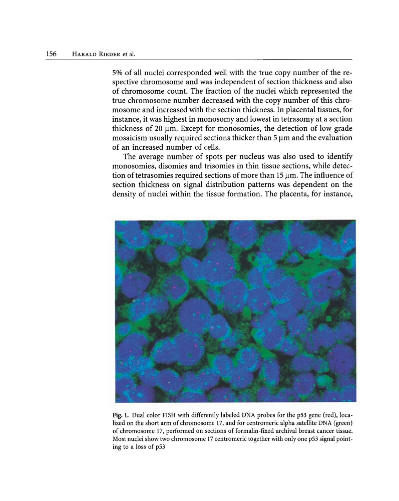
Fig. 1. Dual color FISH with differently labeled DNA probes for the p53 gene (red), localized on the short arm of chromosome 17, and for centromeric alpha satellite DNA (green)
of chromosome 17, performed on sections of formalin-ftxed archival breast cancer tissue.
Most nuclei show two chromosome 17 centromeric together with only one p53 signal pointing to a loss of p53
HARALD RIEDER et al.
5% of all nuclei corresponded well with the true copy number of the respective chromosome and was independent of section thickness and also
of chromosome count. The fraction of the nuclei which represented the
true chromosome number decreased with the copy number of this chromosome and increased with the section thickness. In placental tissues, for
instance, it was highest in monosomy and lowest in tetrasomy at a section
thickness of 20 /lm. Except for monosomies, the detection of low grade
mosaicism usually required sections thicker than 5 /lm and the evaluation
of an increased number of cells.
The average number of spots per nucleus was also used to identify
monosomies, disomies and trisomies in thin tissue sections, while detection of tetrasomies required sections of more than 15 /lm. The influence of
section thickness on signal distribution patterns was dependent on the
density of nuclei within the tissue formation. The placenta, for instance,
Fig. 1. Dual color FISH with differently labeled DNA probes for the p53 gene (red), localized on the short arm of chromosome 17, and for centromeric alpha satellite DNA (green)
of chromosome 17, performed on sections of formalin-ftxed archival breast cancer tissue.
Most nuclei show two chromosome 17 centromeric together with only one p53 signal pointing to a loss of p53