

146
ERICH GEBHART
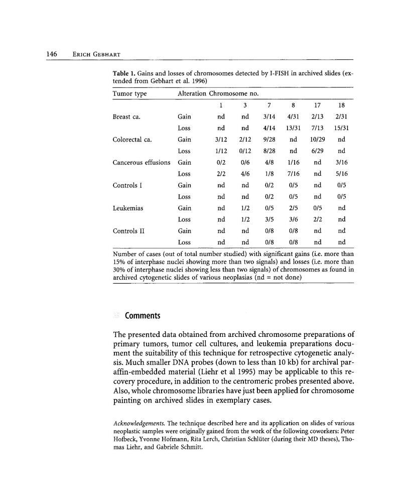
Table 1. Gains and losses of chromosomes detected by I-FISH in archived slides (extended from Gebhart et a1. 1996)
Tumor type
Alteration Chromosome no.
3
7
8
17
18
Breast ca.
Gain
nd
nd
3/14
4/31
2/13
2/31
Loss
nd
nd
4/14
13/31
7/13
15/31
Coloreetal ca.
Gain
3/12
2/12
9/28
nd
10/29
nd
Loss
1/12
0/12
8/28
nd
6/29
nd
Cancerous effusions Gain
0/2
0/6
4/8
1/16
nd
3/16
Loss
2/2
4/6
1/8
7/16
nd
5/16
Controls I
Gain
nd
nd
0/2
0/5
nd
0/5
Loss
nd
nd
0/2
0/5
nd
0/5
Leukemias
Gain
nd
1/2
0/5
2/5
0/5
nd
Loss
nd
1/2
3/5
3/6
2/2
nd
Controls II
Gain
nd
nd
0/8
0/8
nd
nd
Loss
nd
nd
0/8
0/8
nd
nd
Number of cases (out of total number studied) with significant gains (i.e. more than
15% of interphase nuclei showing more than two signals) and losses (i.e. more than
30% of interphase nuclei showing less than two signals) of chromosomes as found in
archived cytogenetic slides of various neoplasias (nd =not done)
Comments
The presented data obtained from archived chromosome preparations of
primary tumors, tumor cell cultures, and leukemia preparations document the suitability of this technique for retrospective cytogenetic analysis. Much smaller DNA probes (down to less than 10 kb) for archival paraffin-embedded material (Liehr et al 1995) may be applicable to this recovery procedure, in addition to the centromeric probes presented above.
Also, whole chromosome libraries have just been applied for chromosome
painting on archived slides in exemplary cases.
Acknowledgements. The technique described here and its application on slides of various
neoplastic samples were originally gained from the work of the following coworkers: Peter
Hotbeck, Yvonne Hofmann, Rita Lerch, Christian Schliiter (during their MD theses), Thomas Liehr, and Gabriele Schmitt.
ERICH GEBHART
Table 1. Gains and losses of chromosomes detected by I-FISH in archived slides (extended from Gebhart et a1. 1996)
Tumor type
Alteration Chromosome no.
3
7
8
17
18
Breast ca.
Gain
nd
nd
3/14
4/31
2/13
2/31
Loss
nd
nd
4/14
13/31
7/13
15/31
Coloreetal ca.
Gain
3/12
2/12
9/28
nd
10/29
nd
Loss
1/12
0/12
8/28
nd
6/29
nd
Cancerous effusions Gain
0/2
0/6
4/8
1/16
nd
3/16
Loss
2/2
4/6
1/8
7/16
nd
5/16
Controls I
Gain
nd
nd
0/2
0/5
nd
0/5
Loss
nd
nd
0/2
0/5
nd
0/5
Leukemias
Gain
nd
1/2
0/5
2/5
0/5
nd
Loss
nd
1/2
3/5
3/6
2/2
nd
Controls II
Gain
nd
nd
0/8
0/8
nd
nd
Loss
nd
nd
0/8
0/8
nd
nd
Number of cases (out of total number studied) with significant gains (i.e. more than
15% of interphase nuclei showing more than two signals) and losses (i.e. more than
30% of interphase nuclei showing less than two signals) of chromosomes as found in
archived cytogenetic slides of various neoplasias (nd =not done)
Comments
The presented data obtained from archived chromosome preparations of
primary tumors, tumor cell cultures, and leukemia preparations document the suitability of this technique for retrospective cytogenetic analysis. Much smaller DNA probes (down to less than 10 kb) for archival paraffin-embedded material (Liehr et al 1995) may be applicable to this recovery procedure, in addition to the centromeric probes presented above.
Also, whole chromosome libraries have just been applied for chromosome
painting on archived slides in exemplary cases.
Acknowledgements. The technique described here and its application on slides of various
neoplastic samples were originally gained from the work of the following coworkers: Peter
Hotbeck, Yvonne Hofmann, Rita Lerch, Christian Schliiter (during their MD theses), Thomas Liehr, and Gabriele Schmitt.