

10 Archival Cytogenetic Slides
145
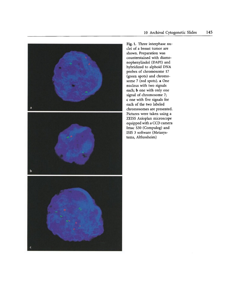
Fig. 1. Three interphase nuclei of a breast tumor are
shown. Preparation was
counterstained with diamonophenylindol (DAPI) and
hybridized to alphoid DNA
probes of chromosome 17
(green spots) and chromosome 7 (red spots). a One
nucleus with two signals
each; b one with only one
signal of chromosome 7;
c one with five signals for
each of the two labeled
chromosomes are presented.
Pictures were taken using a
ZEISS Axioplan microscope
equipped with a CCD camera
Imac S30 (Compulog) and
ISIS 3 software (Metasysterns, Altlussheim)
145
Fig. 1. Three interphase nuclei of a breast tumor are
shown. Preparation was
counterstained with diamonophenylindol (DAPI) and
hybridized to alphoid DNA
probes of chromosome 17
(green spots) and chromosome 7 (red spots). a One
nucleus with two signals
each; b one with only one
signal of chromosome 7;
c one with five signals for
each of the two labeled
chromosomes are presented.
Pictures were taken using a
ZEISS Axioplan microscope
equipped with a CCD camera
Imac S30 (Compulog) and
ISIS 3 software (Metasysterns, Altlussheim)