

220
M.A. Masini et al.
Distribution of ANP Specific Binding Sites
a-rANP binding sites were examined by in vitro autoradiography of Bouinfixed sections using 79 pM 125 1 ANP (specific activity 1397 Ci/mmol,
Peninsula) LM-l emulsion (Amersham, Buckinghamshire, UK) [22].
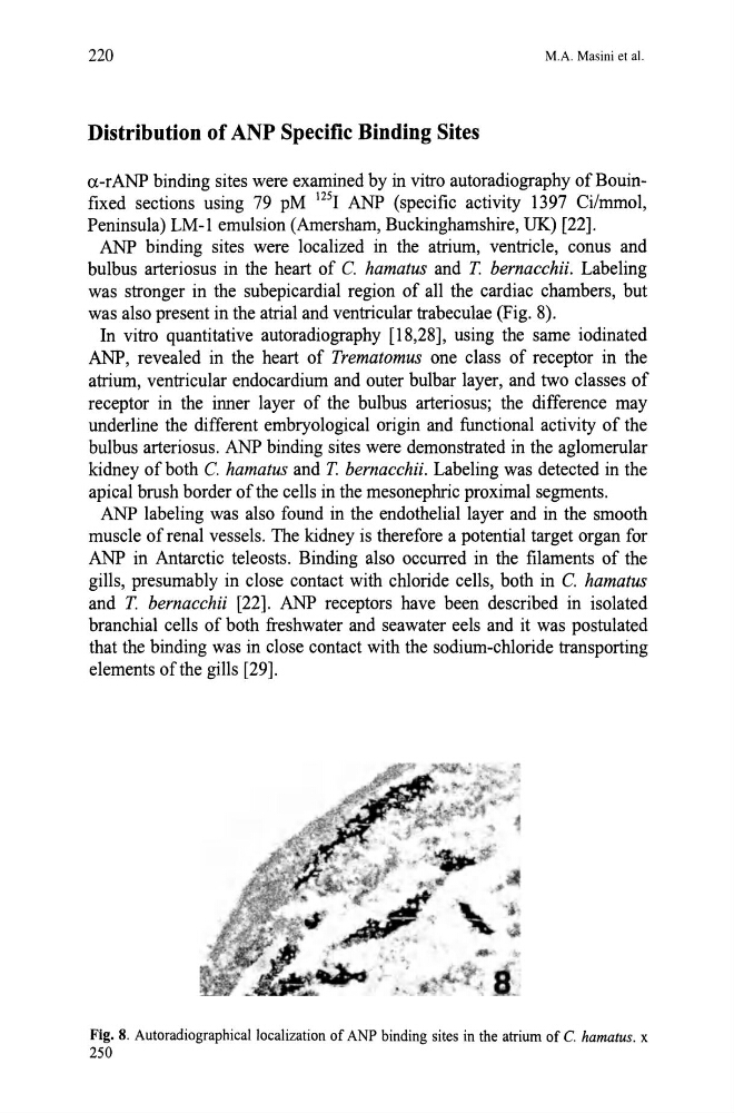
ANP binding sites were localized in the atrium, ventricle, conus and
bulbus arteriosus in the heart of C. hamatus and T. bernacchii. Labeling
was stronger in the subepicardial region of all the cardiac chambers, but
was also present in the atrial and ventricular trabeculae (Fig. 8).
In vitro quantitative autoradiography [18,28], using the same iodinated
ANP, revealed in the heart of Trematomus one class of receptor in the
atrium, ventricular endocardium and outer bulbar layer, and two classes of
receptor in the inner layer of the bulbus arteriosus; the difference may
underline the different embryological origin and functional activity of the
bulbus arteriosus. ANP binding sites were demonstrated in the aglomerular
kidney of both C. hamatus and T. bernacchii. Labeling was detected in the
apical brush border of the cells in the mesonephric proximal segments.
ANP labeling was also found in the endothelial layer and in the smooth
muscle of renal vessels. The kidney is therefore a potential target organ for
ANP in Antarctic teleosts. Binding also occurred in the filaments of the
gills, presumably in close contact with chloride cells, both in C. hamatus
and T. bernacchii [22]. ANP receptors have been described in isolated
branchial cells of both freshwater and seawater eels and it was postulated
that the binding was in close contact with the sodium-chloride transporting
elements of the gills [29].
Fig. 8. Autoradiographicallocalization of ANP binding sites in the atrium of C. hamatus. x
250
M.A. Masini et al.
Distribution of ANP Specific Binding Sites
a-rANP binding sites were examined by in vitro autoradiography of Bouinfixed sections using 79 pM 125 1 ANP (specific activity 1397 Ci/mmol,
Peninsula) LM-l emulsion (Amersham, Buckinghamshire, UK) [22].
ANP binding sites were localized in the atrium, ventricle, conus and
bulbus arteriosus in the heart of C. hamatus and T. bernacchii. Labeling
was stronger in the subepicardial region of all the cardiac chambers, but
was also present in the atrial and ventricular trabeculae (Fig. 8).
In vitro quantitative autoradiography [18,28], using the same iodinated
ANP, revealed in the heart of Trematomus one class of receptor in the
atrium, ventricular endocardium and outer bulbar layer, and two classes of
receptor in the inner layer of the bulbus arteriosus; the difference may
underline the different embryological origin and functional activity of the
bulbus arteriosus. ANP binding sites were demonstrated in the aglomerular
kidney of both C. hamatus and T. bernacchii. Labeling was detected in the
apical brush border of the cells in the mesonephric proximal segments.
ANP labeling was also found in the endothelial layer and in the smooth
muscle of renal vessels. The kidney is therefore a potential target organ for
ANP in Antarctic teleosts. Binding also occurred in the filaments of the
gills, presumably in close contact with chloride cells, both in C. hamatus
and T. bernacchii [22]. ANP receptors have been described in isolated
branchial cells of both freshwater and seawater eels and it was postulated
that the binding was in close contact with the sodium-chloride transporting
elements of the gills [29].
Fig. 8. Autoradiographicallocalization of ANP binding sites in the atrium of C. hamatus. x
250