

Vascular Adaptations for Blood Supply to Locomotory Muscles of Icefish
189
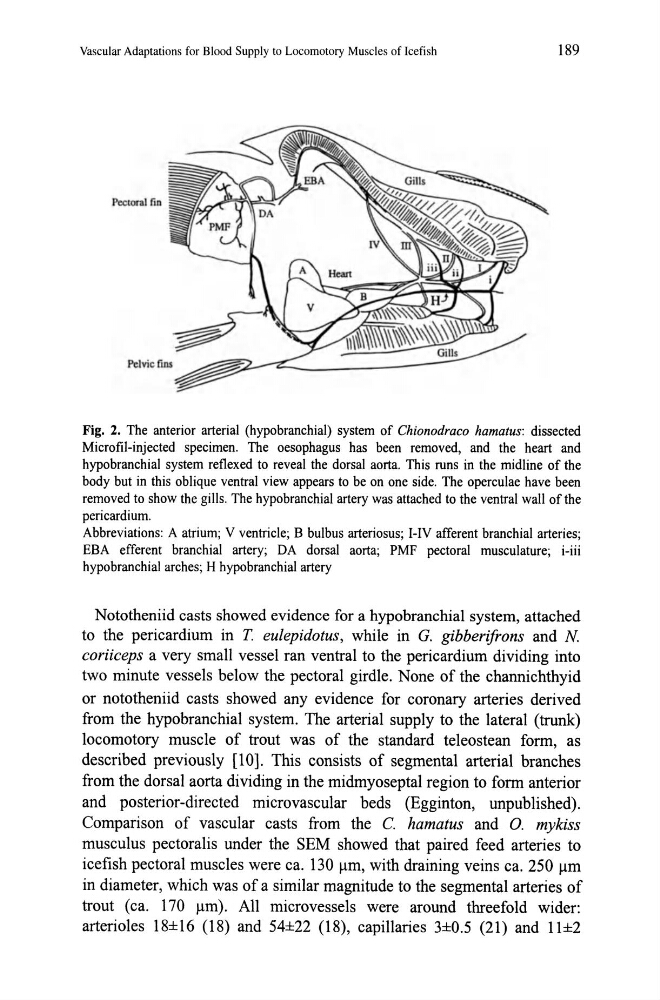
Fig. 2. The anterior arterial (hypobranchial) system of Chionodraco hamatus: dissected
Microfil-injected specimen. The oesophagus has been removed, and the heart and
hypobranchial system reflexed to reveal the dorsal aorta. This runs in the midline of the
body but in this oblique ventral view appears to be on one side. The operculae have been
removed to show the gills. The hypobranchial artery was attached to the ventral wall of the
pericardium.
Abbreviations: A atrium; V ventricle; B bulbus arteriosus; I-IV afferent branchial arteries;
EBA efferent branchial artery; DA dorsal aorta; PMF pectoral musculature; i-iii
hypobranchial arches; H hypobranchial artery
Nototheniid casts showed evidence for a hypobranchial system, attached
to the pericardium in T. eulepidotus, while in G. gibberifrons and N
coriiceps a very small vessel ran ventral to the pericardium dividing into
two minute vessels below the pectoral girdle. None of the channichthyid
or nototheniid casts showed any evidence for coronary arteries derived
from the hypobranchial system. The arterial supply to the lateral (trunk)
locomotory muscle of trout was of the standard teleostean form, as
described previously [10]. This consists of segmental arterial branches
from the dorsal aorta dividing in the midmyoseptal region to form anterior
and posterior-directed microvascular beds (Egginton, unpublished).
Comparison of vascular casts from the C. hamatus and 0. mykiss
musculus pectoralis under the SEM showed that paired feed arteries to
icefish pectoral muscles were ca. 130 Ilm, with draining veins ca. 250 Ilm
in diameter, which was of a similar magnitude to the segmental arteries of
trout (ca. 170 Ilm) . All microvessels were around threefold wider:
arterioles 18±16 (18) and 54±22 (18), capillaries 3±0.5 (21) and 11±2
189
Fig. 2. The anterior arterial (hypobranchial) system of Chionodraco hamatus: dissected
Microfil-injected specimen. The oesophagus has been removed, and the heart and
hypobranchial system reflexed to reveal the dorsal aorta. This runs in the midline of the
body but in this oblique ventral view appears to be on one side. The operculae have been
removed to show the gills. The hypobranchial artery was attached to the ventral wall of the
pericardium.
Abbreviations: A atrium; V ventricle; B bulbus arteriosus; I-IV afferent branchial arteries;
EBA efferent branchial artery; DA dorsal aorta; PMF pectoral musculature; i-iii
hypobranchial arches; H hypobranchial artery
Nototheniid casts showed evidence for a hypobranchial system, attached
to the pericardium in T. eulepidotus, while in G. gibberifrons and N
coriiceps a very small vessel ran ventral to the pericardium dividing into
two minute vessels below the pectoral girdle. None of the channichthyid
or nototheniid casts showed any evidence for coronary arteries derived
from the hypobranchial system. The arterial supply to the lateral (trunk)
locomotory muscle of trout was of the standard teleostean form, as
described previously [10]. This consists of segmental arterial branches
from the dorsal aorta dividing in the midmyoseptal region to form anterior
and posterior-directed microvascular beds (Egginton, unpublished).
Comparison of vascular casts from the C. hamatus and 0. mykiss
musculus pectoralis under the SEM showed that paired feed arteries to
icefish pectoral muscles were ca. 130 Ilm, with draining veins ca. 250 Ilm
in diameter, which was of a similar magnitude to the segmental arteries of
trout (ca. 170 Ilm) . All microvessels were around threefold wider:
arterioles 18±16 (18) and 54±22 (18), capillaries 3±0.5 (21) and 11±2