

188
S. Egginton and J.e. Rankin
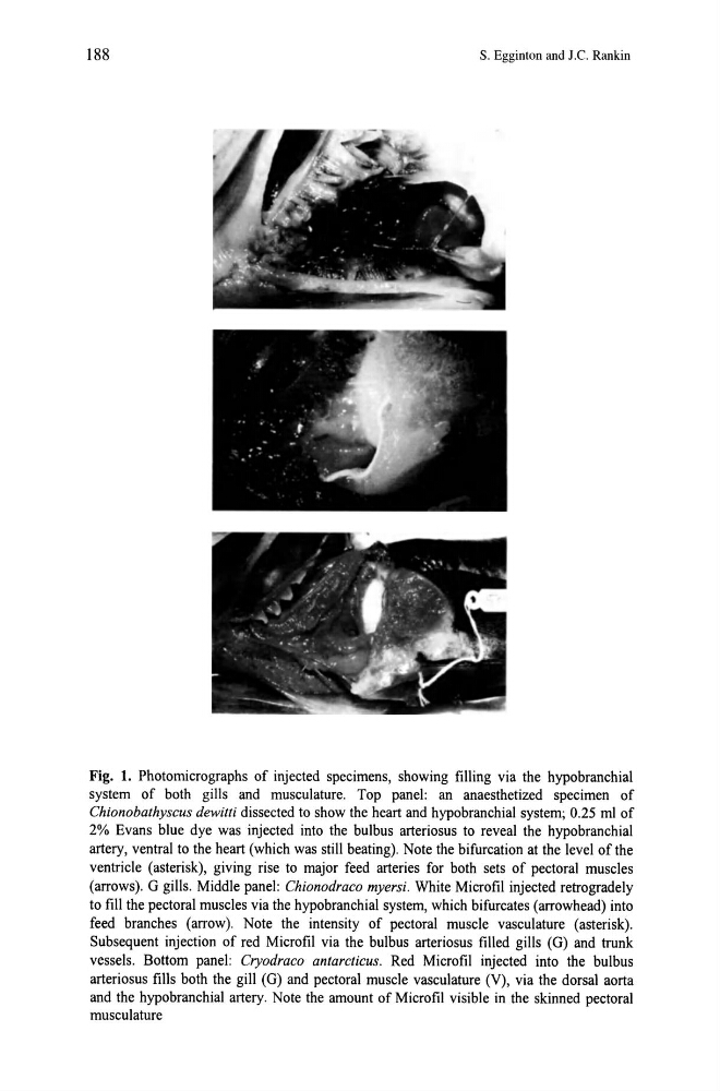
Fig. 1. Photomicrographs of injected specimens, showing filling via the hypobranchial
system of both gills and musculature. Top panel: an anaesthetized specimen of
Chionobathyscus dewitti dissected to show the heart and hypobranchial system; 0.25 ml of
2% Evans blue dye was injected into the bulbus arteriosus to reveal the hypobranchial
artery, ventral to the heart (which was still beating). Note the bifurcation at the level of the
ventricle (asterisk), giving rise to major feed arteries for both sets of pectoral muscles
(arrows). G gills. Middle panel: Chionodraco myersi. White Microfil injected retrogradely
to fill the pectoral muscles via the hypobranchial system, which bifurcates (arrowhead) into
feed branches (arrow). Note the intensity of pectoral muscle vasculature (asterisk).
Subsequent injection of red Microfil via the bulbus arteriosus filled gills (G) and trunk
vessels. Bottom panel: Cryodraco antarcticus. Red Microfil injected into the bulbus
arteriosus fills both the gill (G) and pectoral muscle vasculature (V), via the dorsal aorta
and the hypobranchial artery. Note the amount of Microfil visible in the skinned pectoral
musculature
S. Egginton and J.e. Rankin
Fig. 1. Photomicrographs of injected specimens, showing filling via the hypobranchial
system of both gills and musculature. Top panel: an anaesthetized specimen of
Chionobathyscus dewitti dissected to show the heart and hypobranchial system; 0.25 ml of
2% Evans blue dye was injected into the bulbus arteriosus to reveal the hypobranchial
artery, ventral to the heart (which was still beating). Note the bifurcation at the level of the
ventricle (asterisk), giving rise to major feed arteries for both sets of pectoral muscles
(arrows). G gills. Middle panel: Chionodraco myersi. White Microfil injected retrogradely
to fill the pectoral muscles via the hypobranchial system, which bifurcates (arrowhead) into
feed branches (arrow). Note the intensity of pectoral muscle vasculature (asterisk).
Subsequent injection of red Microfil via the bulbus arteriosus filled gills (G) and trunk
vessels. Bottom panel: Cryodraco antarcticus. Red Microfil injected into the bulbus
arteriosus fills both the gill (G) and pectoral muscle vasculature (V), via the dorsal aorta
and the hypobranchial artery. Note the amount of Microfil visible in the skinned pectoral
musculature