

212
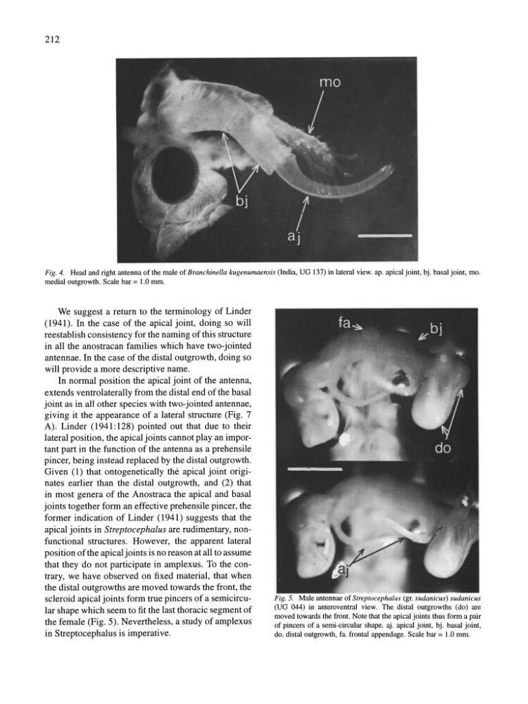
Fig. 4. Head and right antenna of the male of Branchinella kugenumaensis (India, UG 137) in lateral view. ap. apical joint, bj. basal joint, mo.
medial outgrowth. Scale bar = 1.0 mm.
We suggest a return to the terminology of Linder
(1941). In the case of the apical joint, doing so will
reestablish consistency for the naming of this structure
in all the anostracan families which have two-jointed
antennae. In the case of the distal outgrowth, doing so
will provide a more descriptive name.
In normal position the apical joint of the antenna,
extends ventrolaterally from the distal end of the basal
joint as in all other species with two-jointed antennae,
giving it the appearance of a lateral structure (Fig. 7
A). Linder (1941:128) pointed out that due to their
lateral position, the apical joints cannot play an important part in the function of the antenna as a prehensile
pincer, being instead replaced by the distal outgrowth.
Given (1) that ontogenetically the apical joint originates earlier than the distal outgrowth, and (2) that
in most genera of the Anostraca the apical and basal
joints together form an effective prehensile pincer, the
former indication of Linder (1941) suggests that the
apical joints in Streptocephaius are rudimentary, nonfunctional structures. However, the apparent lateral
position of the apical joints is no reason at all to assume
that they do not participate in amplexus. To the contrary, we have observed on fixed material, that when
the distal outgrowths are moved towards the front, the
scleroid apical joints form true pincers of a semicircular shape which seem to fit the last thoracic segment of
the female (Fig. 5). Nevertheless, a study of amplexus
in Streptocephalus is imperative.
Fig. 5. Male antennae of Streptocephalus (gr. sudanicus) sudanicus
(UG 044) in anteroventral view. The distal outgrowths (do) are
moved towards the front. Note that the apical joints thus form a pair
of pincers of a semi-circular shape. aj. apical joint, bj. basal joint,
do. distal outgrowth, fa. frontal appendage. Scale bar = 1.0 mm.
Fig. 4. Head and right antenna of the male of Branchinella kugenumaensis (India, UG 137) in lateral view. ap. apical joint, bj. basal joint, mo.
medial outgrowth. Scale bar = 1.0 mm.
We suggest a return to the terminology of Linder
(1941). In the case of the apical joint, doing so will
reestablish consistency for the naming of this structure
in all the anostracan families which have two-jointed
antennae. In the case of the distal outgrowth, doing so
will provide a more descriptive name.
In normal position the apical joint of the antenna,
extends ventrolaterally from the distal end of the basal
joint as in all other species with two-jointed antennae,
giving it the appearance of a lateral structure (Fig. 7
A). Linder (1941:128) pointed out that due to their
lateral position, the apical joints cannot play an important part in the function of the antenna as a prehensile
pincer, being instead replaced by the distal outgrowth.
Given (1) that ontogenetically the apical joint originates earlier than the distal outgrowth, and (2) that
in most genera of the Anostraca the apical and basal
joints together form an effective prehensile pincer, the
former indication of Linder (1941) suggests that the
apical joints in Streptocephaius are rudimentary, nonfunctional structures. However, the apparent lateral
position of the apical joints is no reason at all to assume
that they do not participate in amplexus. To the contrary, we have observed on fixed material, that when
the distal outgrowths are moved towards the front, the
scleroid apical joints form true pincers of a semicircular shape which seem to fit the last thoracic segment of
the female (Fig. 5). Nevertheless, a study of amplexus
in Streptocephalus is imperative.
Fig. 5. Male antennae of Streptocephalus (gr. sudanicus) sudanicus
(UG 044) in anteroventral view. The distal outgrowths (do) are
moved towards the front. Note that the apical joints thus form a pair
of pincers of a semi-circular shape. aj. apical joint, bj. basal joint,
do. distal outgrowth, fa. frontal appendage. Scale bar = 1.0 mm.