

(1991) stressed the need for a reinvestigation of this
subject in the whole Order Anostraca. Although this
matter deserves a histological analysis, we observed
that the endopodite appears to recede toward the base
of the appendage and degenerate, while the exopodite
increases in size, forming a two-jointed structure (Fig.
1, A). For the female antenna, Baqai (1963) figured the
insertion of the endopodite muscles on the protopodite.
From his figures, it is clear that this insertion moves
in every advanced stage relatively closer to the base of
the appendage, with an apparent reduction of the protopodite, while externally, the endopodite degenerates
and the basal part of the exopodite increases in size
(Baqai, 1963, Figs 20, 22 & 24).
From the distal end of each basal joint arises a process that terminates in a scleroid, cheliform structure
(hand)(Fig. 1, C-G); this is the distal outgrowth of
Linder (1941:134). Also, from the inner side of the
basal joints, two lobes migrate to a medial position
and fuse basally to form the frontal appendage (Baqai,
1963) (Fig. 1, E-H) (frontal appendage = mamelon
(Waga, 1842), frons appendice (Daday, 1908, 191Oa),
appendix frontalis (Daday, 1910a,b), frontal process
(Barnard, 1924, 1929; Bond, 1934), median lamina
(Creaser, 1930». The basal portions of the basal joints
fuse in such a way that they exhibit some degree of
coalescence (Linder, 1941; 134)(Fig. 1, H).
Thus, the antenna of the adult male is a uniramous,
two-jointed appendage, with a typical distal outgrowth
that terminates in a cheliform structure; the so-called
hand (Fig. 2).
In general, the cylindrical, robust, and dorsally
setose basal joint (Linder, 1941)(= basal segment, part
or section (Sars, 1898, 1899, 1900, 1905), articulus
basalis (Daday, 1908, 191Oa,b), first or basal joint
(Barnard, 1924), Basalglieder (Brtek, 1974» (Figs
1, 2 A, 3, 7 A), is similar in all species, except
that in a number of forms it is ornamented by processes. The slender, scleroid, and non-setose apical
joint (Linder, 1941:126)(= claw-like process or spine
(Sars, 1898, 1899, 1900, 1905),processusaculeiformis
or antenniformis (Daday, 1908, 191Oa,b), appendage
(Barnard, 1924), appendix (Barnard, 1929), process
(Bond, 1934), Endglied der Antenne (Brtek, 1974),
terminal joint (Brendonck & Belk, 1993; Hamer et al.
1994a,b) inserted just above the ventral border of the
distal end of the basal joint, is ventrolaterally directed and distally curved inward (Figs 1, 2 A, 5, 7 A,
B). This joint is also similar in all species. Linder
(1941 :231), called this structure the apical joint of a
two-segmented appendage, noting that it represented
211
B
bi
pd
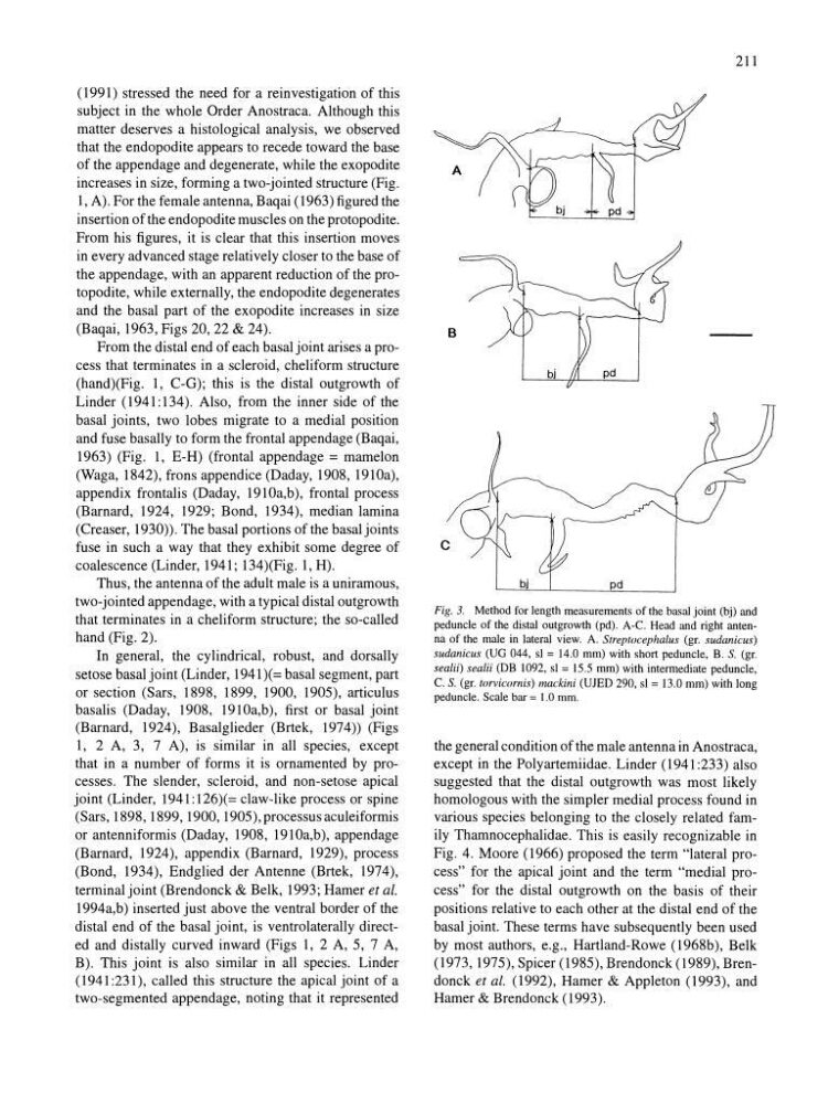
Fig. 3. Method for length measurements of the basal joint (bj) and
peduncle of the distal outgrowtb (pd). A-C. Head and right antenna of the male in lateral view. A. Streptocephaius (gr. sudanicus)
sudanicus (UG 044, sl = 14.0 mm) with short peduncle, B. S. (gr.
sealii) sealii (DB 1092, sl = 15.5 mm) with intermediate peduncle,
C. S. (gr. torvicornis) mackini (UJED 290, sl = 13.0 mm) with long
peduncle. Scale bar = 1.0 mm.
the general condition of the male antenna in Anostraca,
except in the Polyartemiidae. Linder (1941:233) also
suggested that the distal outgrowth was most likely
homologous with the simpler medial process found in
various species belonging to the closely related family Thamnocephalidae. This is easily recognizable in
Fig. 4. Moore (1966) proposed the term "lateral process" for the apical joint and the term "medial process" for the distal outgrowth on the basis of their
positions relative to each other at the distal end of the
basal joint. These terms have subsequently been used
by most authors, e.g., Hartland-Rowe (1968b), Belk
(1973, 1975), Spicer (1985), Brendonck (1989), Brendonck et al. (1992), Hamer & Appleton (1993), and
Hamer & Brendonck (1993).
subject in the whole Order Anostraca. Although this
matter deserves a histological analysis, we observed
that the endopodite appears to recede toward the base
of the appendage and degenerate, while the exopodite
increases in size, forming a two-jointed structure (Fig.
1, A). For the female antenna, Baqai (1963) figured the
insertion of the endopodite muscles on the protopodite.
From his figures, it is clear that this insertion moves
in every advanced stage relatively closer to the base of
the appendage, with an apparent reduction of the protopodite, while externally, the endopodite degenerates
and the basal part of the exopodite increases in size
(Baqai, 1963, Figs 20, 22 & 24).
From the distal end of each basal joint arises a process that terminates in a scleroid, cheliform structure
(hand)(Fig. 1, C-G); this is the distal outgrowth of
Linder (1941:134). Also, from the inner side of the
basal joints, two lobes migrate to a medial position
and fuse basally to form the frontal appendage (Baqai,
1963) (Fig. 1, E-H) (frontal appendage = mamelon
(Waga, 1842), frons appendice (Daday, 1908, 191Oa),
appendix frontalis (Daday, 1910a,b), frontal process
(Barnard, 1924, 1929; Bond, 1934), median lamina
(Creaser, 1930». The basal portions of the basal joints
fuse in such a way that they exhibit some degree of
coalescence (Linder, 1941; 134)(Fig. 1, H).
Thus, the antenna of the adult male is a uniramous,
two-jointed appendage, with a typical distal outgrowth
that terminates in a cheliform structure; the so-called
hand (Fig. 2).
In general, the cylindrical, robust, and dorsally
setose basal joint (Linder, 1941)(= basal segment, part
or section (Sars, 1898, 1899, 1900, 1905), articulus
basalis (Daday, 1908, 191Oa,b), first or basal joint
(Barnard, 1924), Basalglieder (Brtek, 1974» (Figs
1, 2 A, 3, 7 A), is similar in all species, except
that in a number of forms it is ornamented by processes. The slender, scleroid, and non-setose apical
joint (Linder, 1941:126)(= claw-like process or spine
(Sars, 1898, 1899, 1900, 1905),processusaculeiformis
or antenniformis (Daday, 1908, 191Oa,b), appendage
(Barnard, 1924), appendix (Barnard, 1929), process
(Bond, 1934), Endglied der Antenne (Brtek, 1974),
terminal joint (Brendonck & Belk, 1993; Hamer et al.
1994a,b) inserted just above the ventral border of the
distal end of the basal joint, is ventrolaterally directed and distally curved inward (Figs 1, 2 A, 5, 7 A,
B). This joint is also similar in all species. Linder
(1941 :231), called this structure the apical joint of a
two-segmented appendage, noting that it represented
211
B
bi
pd
Fig. 3. Method for length measurements of the basal joint (bj) and
peduncle of the distal outgrowtb (pd). A-C. Head and right antenna of the male in lateral view. A. Streptocephaius (gr. sudanicus)
sudanicus (UG 044, sl = 14.0 mm) with short peduncle, B. S. (gr.
sealii) sealii (DB 1092, sl = 15.5 mm) with intermediate peduncle,
C. S. (gr. torvicornis) mackini (UJED 290, sl = 13.0 mm) with long
peduncle. Scale bar = 1.0 mm.
the general condition of the male antenna in Anostraca,
except in the Polyartemiidae. Linder (1941:233) also
suggested that the distal outgrowth was most likely
homologous with the simpler medial process found in
various species belonging to the closely related family Thamnocephalidae. This is easily recognizable in
Fig. 4. Moore (1966) proposed the term "lateral process" for the apical joint and the term "medial process" for the distal outgrowth on the basis of their
positions relative to each other at the distal end of the
basal joint. These terms have subsequently been used
by most authors, e.g., Hartland-Rowe (1968b), Belk
(1973, 1975), Spicer (1985), Brendonck (1989), Brendonck et al. (1992), Hamer & Appleton (1993), and
Hamer & Brendonck (1993).