

134
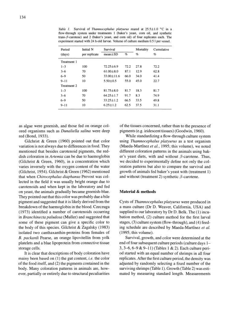
Table 1. Survival of Thamnocephalus platyurus reared at 25.5±1.0 °C in a
flow-through system under treatments I (baker's yeast, corn oil, and synthetic
trans-,6-carotene) and 2 (baker's yeast, and corn oil) of four replicates each. The
experiment started with 24 h-old larvae. Volume of culture medium 0.5 I per vessel.
Period
Initial N
Survival
Mortality
Cumulative
(days)
per replicate
mean±SD
%
%
%
Treatment I
1-3
100
72.25±6.9
72.2 27.8
72.2
3-6
70
61.00±8.0
87.1
12.9
62.8
6-9
50
33.00±I1.6 66.0 34.0
41.4
9-11
10
5.50±0.5
Treatment 2
1-3
100
81.75±8.0
3-6
70
64.25±1.7
6-9
50
33.25±1.2
9-11
10
6.25±1.2
as algae were greenish, and those fed on orange colored organisms such as Dunaliella salina were deep
red (Bond, 1933).
Gilchrist & Green (1960) pointed out that color
variation is not always due to differences in food. They
mentioned that besides carotenoid pigments, the reddish coloration in Artemia can be due to haemoglobin
(Gilchrist & Green, 1960), in a concentration which
varies inversely with the oxygen content of the water
(Gilchrist, 1954). Gilchrist & Green (1962) mentioned
that when Chirocephalus diaphanus Prevost was collected in the field it was usually bright orange due to
carotenoids and when kept in the laboratory and fed
on yeast, the animals gradually became greenish-blue.
They pointed out that this color was probably due a bile
pigment and suggested that it is likely derived from the
breakdown of the haemoglobin in the blood. Czeczuga
(1973) identified a number of carotenoids occurring
in Branchinecta paludosa (Muller) and suggested that
some of these pigment can give a specific color to
the body of this species. Gilchrist & Zagalsky (1983)
isolated two canthaxanthin-proteins from females of
B. packardi Pearse, an orange lipovitellin from yolk
platelets and a blue lipoprotein from connective tissue
storage cells.
It is clear that descriptions of body coloration have
mainy been based on (1) the gut content, i.e. the color
of the food itself, and (2) the pigments contained in the
body. Many coloration patterns in animals are, however, partially or entirely due to structural peculiarities
55.0 45.0
22.7
81.7
18.3
81.7
91.7
8.3
74.9
66.5
33.5
49.8
62.5
37.5
31.1
of the tissues concerned, rather than to the presence of
pigments (e.g. iridescenttissues) (Goodwin, 1960).
While standardizing a flow-through culture system
using Thamnocephalus platyurus as a test organism
(Maeda-Martinez et al., 1995; this volume), we noted
different coloration patterns in the animals using baker's yeast diets, with and without f1-carotene. Thus,
we decided to experimentally define not only the coloration patterns but also to compare the survival and
growth of animals fed baker's yeast with (treatment 1)
and without (treatment 2) synthetic f1-carotene.
Material & methods
Cysts of Thamnocephalus platyurus were produced in
a mass culture (Dr D. Weaver, California, USA) and
supplied to our laboratory by Dr D. Belk. The (1) incubation method, (2) culture method for the first larval
stages, (3) culture system (flow-through), and (4) feeding schedule are described by Maeda-Martinez et at.
(1995; this volume).
Survival, growth, and color were determined at the
end of four subsequent culture periods (culture days 13, 3-6, 6-9 & 9-11) (Tables 1 & 2). Each culture period started with an equal number of shrimps in all four
replicates. After the first culture period, the density was
adjusted by randomly selecting a fixed number of the
surviving shrimps (Table 1). Growth (Table 2) was estimated by measuring standard length. Measurements
Table 1. Survival of Thamnocephalus platyurus reared at 25.5±1.0 °C in a
flow-through system under treatments I (baker's yeast, corn oil, and synthetic
trans-,6-carotene) and 2 (baker's yeast, and corn oil) of four replicates each. The
experiment started with 24 h-old larvae. Volume of culture medium 0.5 I per vessel.
Period
Initial N
Survival
Mortality
Cumulative
(days)
per replicate
mean±SD
%
%
%
Treatment I
1-3
100
72.25±6.9
72.2 27.8
72.2
3-6
70
61.00±8.0
87.1
12.9
62.8
6-9
50
33.00±I1.6 66.0 34.0
41.4
9-11
10
5.50±0.5
Treatment 2
1-3
100
81.75±8.0
3-6
70
64.25±1.7
6-9
50
33.25±1.2
9-11
10
6.25±1.2
as algae were greenish, and those fed on orange colored organisms such as Dunaliella salina were deep
red (Bond, 1933).
Gilchrist & Green (1960) pointed out that color
variation is not always due to differences in food. They
mentioned that besides carotenoid pigments, the reddish coloration in Artemia can be due to haemoglobin
(Gilchrist & Green, 1960), in a concentration which
varies inversely with the oxygen content of the water
(Gilchrist, 1954). Gilchrist & Green (1962) mentioned
that when Chirocephalus diaphanus Prevost was collected in the field it was usually bright orange due to
carotenoids and when kept in the laboratory and fed
on yeast, the animals gradually became greenish-blue.
They pointed out that this color was probably due a bile
pigment and suggested that it is likely derived from the
breakdown of the haemoglobin in the blood. Czeczuga
(1973) identified a number of carotenoids occurring
in Branchinecta paludosa (Muller) and suggested that
some of these pigment can give a specific color to
the body of this species. Gilchrist & Zagalsky (1983)
isolated two canthaxanthin-proteins from females of
B. packardi Pearse, an orange lipovitellin from yolk
platelets and a blue lipoprotein from connective tissue
storage cells.
It is clear that descriptions of body coloration have
mainy been based on (1) the gut content, i.e. the color
of the food itself, and (2) the pigments contained in the
body. Many coloration patterns in animals are, however, partially or entirely due to structural peculiarities
55.0 45.0
22.7
81.7
18.3
81.7
91.7
8.3
74.9
66.5
33.5
49.8
62.5
37.5
31.1
of the tissues concerned, rather than to the presence of
pigments (e.g. iridescenttissues) (Goodwin, 1960).
While standardizing a flow-through culture system
using Thamnocephalus platyurus as a test organism
(Maeda-Martinez et al., 1995; this volume), we noted
different coloration patterns in the animals using baker's yeast diets, with and without f1-carotene. Thus,
we decided to experimentally define not only the coloration patterns but also to compare the survival and
growth of animals fed baker's yeast with (treatment 1)
and without (treatment 2) synthetic f1-carotene.
Material & methods
Cysts of Thamnocephalus platyurus were produced in
a mass culture (Dr D. Weaver, California, USA) and
supplied to our laboratory by Dr D. Belk. The (1) incubation method, (2) culture method for the first larval
stages, (3) culture system (flow-through), and (4) feeding schedule are described by Maeda-Martinez et at.
(1995; this volume).
Survival, growth, and color were determined at the
end of four subsequent culture periods (culture days 13, 3-6, 6-9 & 9-11) (Tables 1 & 2). Each culture period started with an equal number of shrimps in all four
replicates. After the first culture period, the density was
adjusted by randomly selecting a fixed number of the
surviving shrimps (Table 1). Growth (Table 2) was estimated by measuring standard length. Measurements