

121
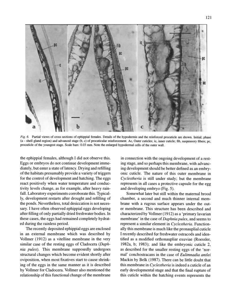
Fig. 6. Partial views of cross sections of ephippial females . Details of the hypodermis and the reinforced procuticle are shown. Initial; phase
(a - shell gland region) and advanced stage (b, c) of procutieular reinforcement. Ac, Outer cuticles; ie, inner cuticle; fib, suspensory fibers; pc,
procutic1e of the youngest stage. Scale bars: 0.03 mm. Note the enlarged hypodermal cells of the outer wall.
the ephippial females, although I did not observe this.
Eggs or embryos do not continue development immediately, but enter a state of latency. Drying and refilling
of the habitats presumably provide a variety of triggers
for the control of development and hatching. The eggs
react positively when water temperature and conductivity levels change, as for example, after heavy rainfall. Laboratory experiments corroborate this. Typically, development restarts after drought and refilling of
the ponds. Nevertheless, total desiccation is not necessary. I have often observed ephippial eggs developing
after filling of only partially dried freshwater bodies. In
these cases, the eggs had remained completely hydrated during the rainless period.
The recently deposited ephippial eggs are enclosed
in an external membrane which was described by
Vollmer (1912) as a vitelline membrane in the very
similar case of the resting eggs of Cladocera (Daphnia pulex). This membrane supposedly undergoes
structural changes which become evident shortly after
oviposition, when most fixatives start to cause shrinking of the eggs in the same manner as it is described
by Vollmer for Cladocera. Vollmer also mentioned the
relationship of this functional change of the membrane
in connection with the ongoing development of a resting stage, and so perhaps this membrane, with advancing development should be better defined as an embryonic cuticle. The nature of this outer membrane in
Cyclestheria is still under study; but the membrane
represents in all cases a protective capsule for the egg
and developing embryo (Fig. 5).
Somewhat later but still within the maternal brood
chamber, a second and much thinner internal membrane with a rugous surface appears under the outer membrane. This structure has been described and
characterized by Vollmer (1912) as a 'primary lavarian
membrane' in the case of Daphnia pulex, and seems to
represent a similar element in Cyclestheria. Functionally this membrane is much like the pronauplial cuticle
I recently described for freshwater ostracods and identified as a modified orthonaupliar exuviae (Roessler,
1982a, b; 1983); and like the embryonic cuticle 2,
as described for the smaller resting eggs of the 'normal' conchostracans in the case of Eulimnadia antlei
Mackin by Belk (1987). There can be little doubt that
this membrane in Cyclestheria is indeed a cuticle of an
early developmental stage and that the final rupture of
this cuticle within the hatching events represents the
Fig. 6. Partial views of cross sections of ephippial females . Details of the hypodermis and the reinforced procuticle are shown. Initial; phase
(a - shell gland region) and advanced stage (b, c) of procutieular reinforcement. Ac, Outer cuticles; ie, inner cuticle; fib, suspensory fibers; pc,
procutic1e of the youngest stage. Scale bars: 0.03 mm. Note the enlarged hypodermal cells of the outer wall.
the ephippial females, although I did not observe this.
Eggs or embryos do not continue development immediately, but enter a state of latency. Drying and refilling
of the habitats presumably provide a variety of triggers
for the control of development and hatching. The eggs
react positively when water temperature and conductivity levels change, as for example, after heavy rainfall. Laboratory experiments corroborate this. Typically, development restarts after drought and refilling of
the ponds. Nevertheless, total desiccation is not necessary. I have often observed ephippial eggs developing
after filling of only partially dried freshwater bodies. In
these cases, the eggs had remained completely hydrated during the rainless period.
The recently deposited ephippial eggs are enclosed
in an external membrane which was described by
Vollmer (1912) as a vitelline membrane in the very
similar case of the resting eggs of Cladocera (Daphnia pulex). This membrane supposedly undergoes
structural changes which become evident shortly after
oviposition, when most fixatives start to cause shrinking of the eggs in the same manner as it is described
by Vollmer for Cladocera. Vollmer also mentioned the
relationship of this functional change of the membrane
in connection with the ongoing development of a resting stage, and so perhaps this membrane, with advancing development should be better defined as an embryonic cuticle. The nature of this outer membrane in
Cyclestheria is still under study; but the membrane
represents in all cases a protective capsule for the egg
and developing embryo (Fig. 5).
Somewhat later but still within the maternal brood
chamber, a second and much thinner internal membrane with a rugous surface appears under the outer membrane. This structure has been described and
characterized by Vollmer (1912) as a 'primary lavarian
membrane' in the case of Daphnia pulex, and seems to
represent a similar element in Cyclestheria. Functionally this membrane is much like the pronauplial cuticle
I recently described for freshwater ostracods and identified as a modified orthonaupliar exuviae (Roessler,
1982a, b; 1983); and like the embryonic cuticle 2,
as described for the smaller resting eggs of the 'normal' conchostracans in the case of Eulimnadia antlei
Mackin by Belk (1987). There can be little doubt that
this membrane in Cyclestheria is indeed a cuticle of an
early developmental stage and that the final rupture of
this cuticle within the hatching events represents the