

118
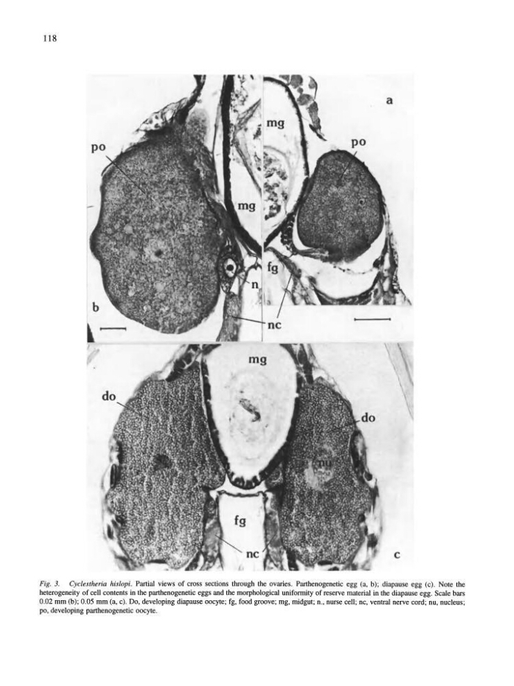
Fig. 3. Cyclestheria hisiopi. Partial views of cross sections through the ovaries. Parthenogenetic egg (a, b); diapause egg (c). Note the
heterogeneity of cell contents in the parthenogenetic eggs and the morphological uniformity of reserve material in the diapause egg. Scale bars
0.02 mm (b); 0.05 mm (a, c). Do, developing diapause oocyte; fg, food groove; mg, midgut; n., nurse cell; nc, ventral nerve cord; nu, nucleus;
po, developing parthenogenetic oocyte.
Fig. 3. Cyclestheria hisiopi. Partial views of cross sections through the ovaries. Parthenogenetic egg (a, b); diapause egg (c). Note the
heterogeneity of cell contents in the parthenogenetic eggs and the morphological uniformity of reserve material in the diapause egg. Scale bars
0.02 mm (b); 0.05 mm (a, c). Do, developing diapause oocyte; fg, food groove; mg, midgut; n., nurse cell; nc, ventral nerve cord; nu, nucleus;
po, developing parthenogenetic oocyte.