

20 In Vitro Toxicity and Histopathological Effects Induced in the Mantle. . .
233
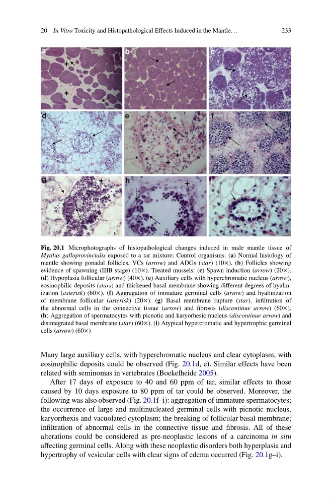
Fig. 20.1 Microphotographs of histopathological changes induced in male mantle tissue of
Mytilus galloprovincialis exposed to a tar mixture: Control organisms: (a) Normal histology of
mantle showing gonadal follicles, VCs (arrow) and ADGs (star) (10). (b) Follicles showing
evidence of spawning (IIIB stage) (10). Treated mussels: (c) Spawn induction (arrow) (20).
(d) Hypoplasia follicular (arrow) (40). (e) Auxiliary cells with hyperchromatic nucleus (arrow),
eosinophilic deposits (stars) and thickened basal membrane showing different degrees of hyalinization (asterisk) (60). (f) Aggregation of immature germinal cells (arrow) and hyalinization
of membrane follicular (asterisk) (20). (g) Basal membrane rupture (star), infiltration of
the abnormal cells in the connective tissue (arrow) and fibrosis (discontinue arrow) (60).
(h) Aggregation of spermatocytes with picnotic and karyorhesic nucleus (discontinue arrow) and
disintegrated basal membrane (star) (60). (i) Atypical hypercromatic and hypertrophic germinal
cells (arrow) (60)
Many large auxiliary cells, with hyperchromatic nucleus and clear cytoplasm, with
eosinophilic deposits could be observed (Fig. 20.1d, e). Similar effects have been
related with seminomas in vertebrates (Boekelheide 2005).
After 17 days of exposure to 40 and 60 ppm of tar, similar effects to those
caused by 10 days exposure to 80 ppm of tar could be observed. Moreover, the
following was also observed (Fig. 20.1f–i): aggregation of immature spermatocytes;
the occurrence of large and multinucleated germinal cells with picnotic nucleus,
karyorrhexis and vacuolated cytoplasm; the breaking of follicular basal membrane;
infiltration of abnormal cells in the connective tissue and fibrosis. All of these
alterations could be considered as pre-neoplastic lesions of a carcinoma in situ
affecting germinal cells. Along with these neoplastic disorders both hyperplasia and
hypertrophy of vesicular cells with clear signs of edema occurred (Fig. 20.1g–i).
233
Fig. 20.1 Microphotographs of histopathological changes induced in male mantle tissue of
Mytilus galloprovincialis exposed to a tar mixture: Control organisms: (a) Normal histology of
mantle showing gonadal follicles, VCs (arrow) and ADGs (star) (10). (b) Follicles showing
evidence of spawning (IIIB stage) (10). Treated mussels: (c) Spawn induction (arrow) (20).
(d) Hypoplasia follicular (arrow) (40). (e) Auxiliary cells with hyperchromatic nucleus (arrow),
eosinophilic deposits (stars) and thickened basal membrane showing different degrees of hyalinization (asterisk) (60). (f) Aggregation of immature germinal cells (arrow) and hyalinization
of membrane follicular (asterisk) (20). (g) Basal membrane rupture (star), infiltration of
the abnormal cells in the connective tissue (arrow) and fibrosis (discontinue arrow) (60).
(h) Aggregation of spermatocytes with picnotic and karyorhesic nucleus (discontinue arrow) and
disintegrated basal membrane (star) (60). (i) Atypical hypercromatic and hypertrophic germinal
cells (arrow) (60)
Many large auxiliary cells, with hyperchromatic nucleus and clear cytoplasm, with
eosinophilic deposits could be observed (Fig. 20.1d, e). Similar effects have been
related with seminomas in vertebrates (Boekelheide 2005).
After 17 days of exposure to 40 and 60 ppm of tar, similar effects to those
caused by 10 days exposure to 80 ppm of tar could be observed. Moreover, the
following was also observed (Fig. 20.1f–i): aggregation of immature spermatocytes;
the occurrence of large and multinucleated germinal cells with picnotic nucleus,
karyorrhexis and vacuolated cytoplasm; the breaking of follicular basal membrane;
infiltration of abnormal cells in the connective tissue and fibrosis. All of these
alterations could be considered as pre-neoplastic lesions of a carcinoma in situ
affecting germinal cells. Along with these neoplastic disorders both hyperplasia and
hypertrophy of vesicular cells with clear signs of edema occurred (Fig. 20.1g–i).