

232
A.A. Martinez et al.
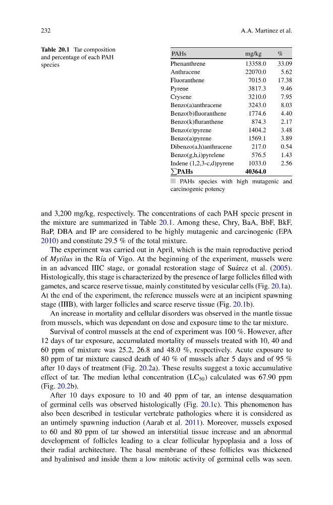
Table 20.1 Tar composition
and percentage of each PAH
species
PAHs
mg/kg
%
Phenanthrene
13358.0
33.09
Anthracene
22070.0
5.62
Fluoranthene
7015.0
17.38
Pyrene
3817.3
9.46
Crysene
3210.0
7.95
Benzo(a)anthracene
3243.0
8.03
Benzo(b)fluoranthene
1774.6
4.40
Benzo(k)fluranthene
874.3
2.17
Benzo(e)pyrene
1404.2
3.48
Benzo(a)pyrene
1569.1
3.89
Dibenzo(a,h)anthracene
217.0
0.54
Benzo(g,h,i)pyrelene
576.5
1.43
Indene (1,2,3-c,d)pyrene
1033.0
2.56
P
PAHs
40364.0
PAHs species with high mutagenic and
carcinogenic potency
and 3,200 mg/kg, respectively. The concentrations of each PAH specie present in
the mixture are summarized in Table 20.1. Among these, Chry, BaA, BbF, BkF,
BaP, DBA and IP are considered to be highly mutagenic and carcinogenic (EPA
2010) and constitute 29.5 % of the total mixture.
The experiment was carried out in April, which is the main reproductive period
of Mytilus in the R´ ıa of Vigo. At the beginning of the experiment, mussels were
in an advanced IIIC stage, or gonadal restoration stage of Su´ arez et al. (2005).
Histologically, this stage is characterized by the presence of large follicles filled with
gametes, and scarce reserve tissue, mainly constituted by vesicular cells (Fig. 20.1a).
At the end of the experiment, the reference mussels were at an incipient spawning
stage (IIIB), with larger follicles and scarce reserve tissue (Fig. 20.1b).
An increase in mortality and cellular disorders was observed in the mantle tissue
from mussels, which was dependant on dose and exposure time to the tar mixture.
Survival of control mussels at the end of experiment was 100 %. However, after
12 days of tar exposure, accumulated mortality of mussels treated with 10, 40 and
60 ppm of mixture was 25.2, 26.8 and 48.0 %, respectively. Acute exposure to
80 ppm of tar mixture caused death of 40 % of mussels after 5 days and of 95 %
after 10 days of treatment (Fig. 20.2a). These results suggest a toxic accumulative
effect of tar. The median lethal concentration (LC 50 ) calculated was 67.90 ppm
(Fig. 20.2b).
After 10 days exposure to 10 and 40 ppm of tar, an intense desquamation
of germinal cells was observed histologically (Fig. 20.1c). This phenomenon has
also been described in testicular vertebrate pathologies where it is considered as
an untimely spawning induction (Aarab et al. 2011). Moreover, mussels exposed
to 60 and 80 ppm of tar showed an interstitial tissue increase and an abnormal
development of follicles leading to a clear follicular hypoplasia and a loss of
their radial architecture. The basal membrane of these follicles was thickened
and hyalinised and inside them a low mitotic activity of germinal cells was seen.
A.A. Martinez et al.
Table 20.1 Tar composition
and percentage of each PAH
species
PAHs
mg/kg
%
Phenanthrene
13358.0
33.09
Anthracene
22070.0
5.62
Fluoranthene
7015.0
17.38
Pyrene
3817.3
9.46
Crysene
3210.0
7.95
Benzo(a)anthracene
3243.0
8.03
Benzo(b)fluoranthene
1774.6
4.40
Benzo(k)fluranthene
874.3
2.17
Benzo(e)pyrene
1404.2
3.48
Benzo(a)pyrene
1569.1
3.89
Dibenzo(a,h)anthracene
217.0
0.54
Benzo(g,h,i)pyrelene
576.5
1.43
Indene (1,2,3-c,d)pyrene
1033.0
2.56
P
PAHs
40364.0
PAHs species with high mutagenic and
carcinogenic potency
and 3,200 mg/kg, respectively. The concentrations of each PAH specie present in
the mixture are summarized in Table 20.1. Among these, Chry, BaA, BbF, BkF,
BaP, DBA and IP are considered to be highly mutagenic and carcinogenic (EPA
2010) and constitute 29.5 % of the total mixture.
The experiment was carried out in April, which is the main reproductive period
of Mytilus in the R´ ıa of Vigo. At the beginning of the experiment, mussels were
in an advanced IIIC stage, or gonadal restoration stage of Su´ arez et al. (2005).
Histologically, this stage is characterized by the presence of large follicles filled with
gametes, and scarce reserve tissue, mainly constituted by vesicular cells (Fig. 20.1a).
At the end of the experiment, the reference mussels were at an incipient spawning
stage (IIIB), with larger follicles and scarce reserve tissue (Fig. 20.1b).
An increase in mortality and cellular disorders was observed in the mantle tissue
from mussels, which was dependant on dose and exposure time to the tar mixture.
Survival of control mussels at the end of experiment was 100 %. However, after
12 days of tar exposure, accumulated mortality of mussels treated with 10, 40 and
60 ppm of mixture was 25.2, 26.8 and 48.0 %, respectively. Acute exposure to
80 ppm of tar mixture caused death of 40 % of mussels after 5 days and of 95 %
after 10 days of treatment (Fig. 20.2a). These results suggest a toxic accumulative
effect of tar. The median lethal concentration (LC 50 ) calculated was 67.90 ppm
(Fig. 20.2b).
After 10 days exposure to 10 and 40 ppm of tar, an intense desquamation
of germinal cells was observed histologically (Fig. 20.1c). This phenomenon has
also been described in testicular vertebrate pathologies where it is considered as
an untimely spawning induction (Aarab et al. 2011). Moreover, mussels exposed
to 60 and 80 ppm of tar showed an interstitial tissue increase and an abnormal
development of follicles leading to a clear follicular hypoplasia and a loss of
their radial architecture. The basal membrane of these follicles was thickened
and hyalinised and inside them a low mitotic activity of germinal cells was seen.