

small intestine and a descending large intestine that connects to the cloaca (DíazMiranda et al. 1995, García-Arrarás et al. 1998). In addition, many of the investigations provide important information on the neuroanatomy of sea cucumbers,
including a description of the nervous system in the digestive tract and tube feet
and the discovery of new nerve plexi within the connective tissue, (García-Arrarás
et al. 2001; Díaz-Balzac et al. 2007, 2010a, b) thus making H. glaberrima a much
needed model for understanding neural connections in holothurians.
Neurobiology
Studies on H. glaberrima have provided important information on the nervous
system. The main focus of these studies has been the development and use of
neural markers that serve to identify and characterize components of the nervous
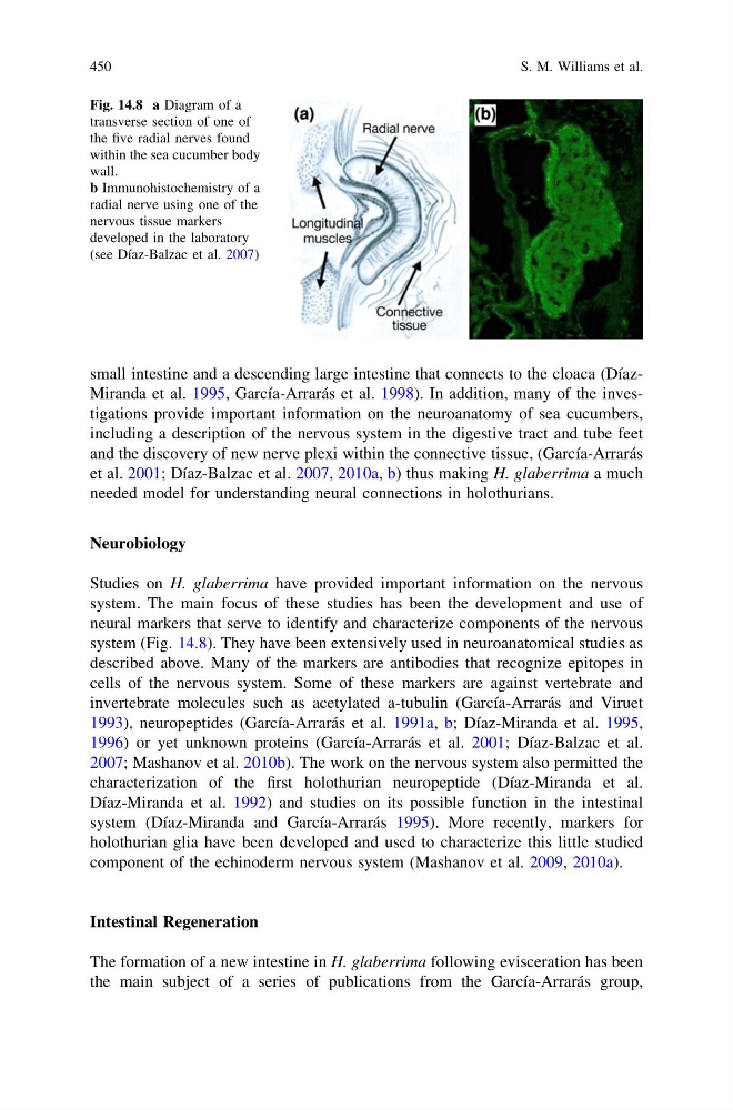
system (Fig. 14.8). They have been extensively used in neuroanatomical studies as
described above. Many of the markers are antibodies that recognize epitopes in
cells of the nervous system. Some of these markers are against vertebrate and
invertebrate molecules such as acetylated a-tubulin (García-Arrarás and Viruet
1993), neuropeptides (García-Arrarás et al. 1991a, b; Díaz-Miranda et al. 1995,
1996) or yet unknown proteins (García-Arrarás et al. 2001; Díaz-Balzac et al.
2007; Mashanov et al. 2010b). The work on the nervous system also permitted the
characterization of the first holothurian neuropeptide (Díaz-Miranda et al.
Díaz-Miranda et al. 1992) and studies on its possible function in the intestinal
system (Díaz-Miranda and García-Arrarás 1995). More recently, markers for
holothurian glia have been developed and used to characterize this little studied
component of the echinoderm nervous system (Mashanov et al. 2009, 2010a).
Intestinal Regeneration
The formation of a new intestine in H. glaberrima following evisceration has been
the main subject of a series of publications from the García-Arrarás group,
Fig. 14.8 a Diagram of a
transverse section of one of
the five radial nerves found
within the sea cucumber body
wall.
b Immunohistochemistry of a
radial nerve using one of the
nervous tissue markers
developed in the laboratory
(see Díaz-Balzac et al. 2007)
450
S. M. Williams et al.
including a description of the nervous system in the digestive tract and tube feet
and the discovery of new nerve plexi within the connective tissue, (García-Arrarás
et al. 2001; Díaz-Balzac et al. 2007, 2010a, b) thus making H. glaberrima a much
needed model for understanding neural connections in holothurians.
Neurobiology
Studies on H. glaberrima have provided important information on the nervous
system. The main focus of these studies has been the development and use of
neural markers that serve to identify and characterize components of the nervous
system (Fig. 14.8). They have been extensively used in neuroanatomical studies as
described above. Many of the markers are antibodies that recognize epitopes in
cells of the nervous system. Some of these markers are against vertebrate and
invertebrate molecules such as acetylated a-tubulin (García-Arrarás and Viruet
1993), neuropeptides (García-Arrarás et al. 1991a, b; Díaz-Miranda et al. 1995,
1996) or yet unknown proteins (García-Arrarás et al. 2001; Díaz-Balzac et al.
2007; Mashanov et al. 2010b). The work on the nervous system also permitted the
characterization of the first holothurian neuropeptide (Díaz-Miranda et al.
Díaz-Miranda et al. 1992) and studies on its possible function in the intestinal
system (Díaz-Miranda and García-Arrarás 1995). More recently, markers for
holothurian glia have been developed and used to characterize this little studied
component of the echinoderm nervous system (Mashanov et al. 2009, 2010a).
Intestinal Regeneration
The formation of a new intestine in H. glaberrima following evisceration has been
the main subject of a series of publications from the García-Arrarás group,
Fig. 14.8 a Diagram of a
transverse section of one of
the five radial nerves found
within the sea cucumber body
wall.
b Immunohistochemistry of a
radial nerve using one of the
nervous tissue markers
developed in the laboratory
(see Díaz-Balzac et al. 2007)
450
S. M. Williams et al.