

72
3. MEASURING GROWTH AND FORM
3.2 Morphological Analysis of a Branching Sponge
In Sect. 2.2.2 the biology of the branching sponge Raspailia inaequalis was
described. Because of its planar form, accurate measurements can be made
from an analysis of photographs, making it a convenient species for the study
of sponge growth. In this section a detailed analysis is made of the branching
pattern of Raspailia inaequalis, as described in Abraham (2001).
A prediction of models of sponge growth is that the branching pattern
should be self-similar. To test whether the branching structure of Raspailia
inaequalis is fractal, 45 specimens were harvested and photographed in the
laboratory. The specimens were taken from a site, known as Leigh Reef,where
Raspailia inaequalis is abundant. The sponges were growing at 25m depth on
a fiat reef and , in order to minimize variation in environmental conditions, all
the specimens were collected from within a circle of 10 m radius. The sponges
were chosen to represent the range of morphologies which was present at
the site, although the smallest sponges « 5cm high) were not collected.
After being harvested the sponges were laid fiat on a black background
and photographed. The digitized images were used for the morphological
analysis.
3.2.1 Image Processing
Some finger sponges have an amorphous lobed morphology, so the branching pattern is difficult to define unambiguously. In contrast, the skeleton of
Raspailia inaequalis is axially condensed, each finger having a spicule bundle at its core. Because of this structure, the photographic images may be
used to obtain a reliable representation of the branching. The method used is
a common procedure in digital analysis. The image is first cleaned by removing any stray light pixels from the dark background. Some of the sponges
were anastomosed: branches that touched while the sponge was growing had
fused together. In order to define the branching, the anastomoses were removed by inserting a line of zero-valued pixels between fused branches. A few
sponges appeared to have been damaged while growing and had a confused
three-dimensional structure. These specimens were not analyzed. After this
preprocessing the images of the sponges were stripped to single lines of
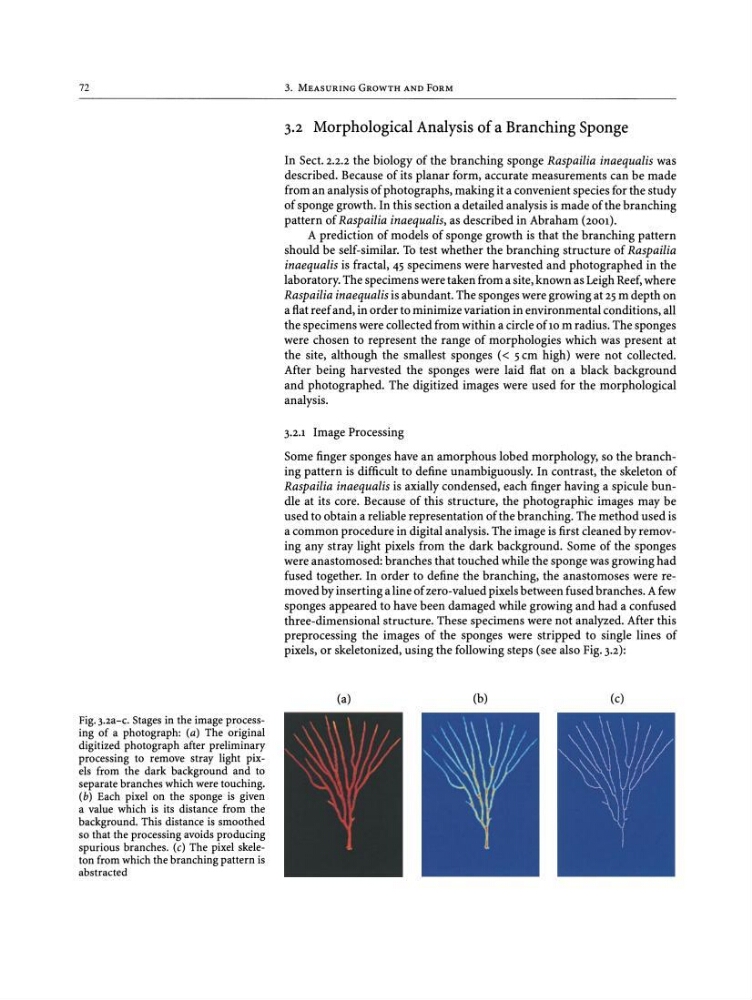
pixels, or skeletonized, using the following steps (see also Fig. 3 .2):
Fig. 3.2a-c. Stages in the imageprocessing of a photograph: (a) The original
digitized photograph after preliminary
processing to remove stray light pixels from the dark background and to
separatebrancheswhichweretouching.
(b) Each pixel on the sponge is given
a value which is its distance from the
background. This distance is smoothed
so that the processing avoids producing
spurious branches. (c) The pixel skeleton fromwhichthe branchingpattern is
abstracted
(a)
(b)
(c)
3. MEASURING GROWTH AND FORM
3.2 Morphological Analysis of a Branching Sponge
In Sect. 2.2.2 the biology of the branching sponge Raspailia inaequalis was
described. Because of its planar form, accurate measurements can be made
from an analysis of photographs, making it a convenient species for the study
of sponge growth. In this section a detailed analysis is made of the branching
pattern of Raspailia inaequalis, as described in Abraham (2001).
A prediction of models of sponge growth is that the branching pattern
should be self-similar. To test whether the branching structure of Raspailia
inaequalis is fractal, 45 specimens were harvested and photographed in the
laboratory. The specimens were taken from a site, known as Leigh Reef,where
Raspailia inaequalis is abundant. The sponges were growing at 25m depth on
a fiat reef and , in order to minimize variation in environmental conditions, all
the specimens were collected from within a circle of 10 m radius. The sponges
were chosen to represent the range of morphologies which was present at
the site, although the smallest sponges « 5cm high) were not collected.
After being harvested the sponges were laid fiat on a black background
and photographed. The digitized images were used for the morphological
analysis.
3.2.1 Image Processing
Some finger sponges have an amorphous lobed morphology, so the branching pattern is difficult to define unambiguously. In contrast, the skeleton of
Raspailia inaequalis is axially condensed, each finger having a spicule bundle at its core. Because of this structure, the photographic images may be
used to obtain a reliable representation of the branching. The method used is
a common procedure in digital analysis. The image is first cleaned by removing any stray light pixels from the dark background. Some of the sponges
were anastomosed: branches that touched while the sponge was growing had
fused together. In order to define the branching, the anastomoses were removed by inserting a line of zero-valued pixels between fused branches. A few
sponges appeared to have been damaged while growing and had a confused
three-dimensional structure. These specimens were not analyzed. After this
preprocessing the images of the sponges were stripped to single lines of
pixels, or skeletonized, using the following steps (see also Fig. 3 .2):
Fig. 3.2a-c. Stages in the imageprocessing of a photograph: (a) The original
digitized photograph after preliminary
processing to remove stray light pixels from the dark background and to
separatebrancheswhichweretouching.
(b) Each pixel on the sponge is given
a value which is its distance from the
background. This distance is smoothed
so that the processing avoids producing
spurious branches. (c) The pixel skeleton fromwhichthe branchingpattern is
abstracted
(a)
(b)
(c)