

2.2. THE CASE STUDIES
59
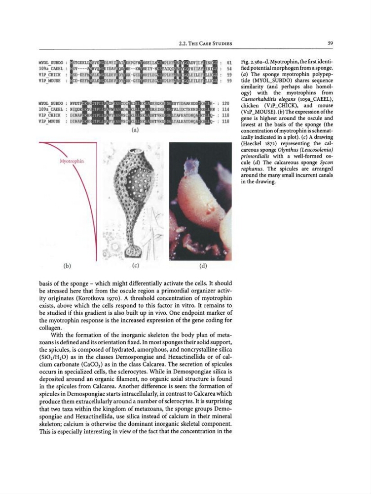
Fig. 2.36a-d. Myotrophin , the first identified potential morphogen from a sponge.
(a) The sponge myotrophin polypeptide (MYOL_SUBDO) shares sequence
similarity (and perhaps also homo logy) with the myotrophins from
Caenorhabditis elegans (109a_CAEEL),
chicken (VIP_CHICK), and mouse
(VIP_MOUSE). (b) The expression of the
gene is highest around the oscule and
lowest at the basis of the sponge (the
concentration of myotrophin is schematically indicated in a plot). (c) A drawing
(Haeckel 1872) representing the calcareous sponge Olynthus (Leucosolenia)
primordialis with a well-formed oscule (d) The calcareous sponge Sycon
raphanus. The spicules are arranged
around the many small incurrent canals
in the drawing.
61
54
59
59
120
114
118
118
STIDAAESD
ALIDC1EEEoD1El!!KlI
TAFEATDNQ
TALEATDNQ
LYEli i GFll
ID AYI S E- - KH
LOEYt
' - GE
LOEY'
- GE
(a)
Myotrophin
HYOL_SUBOO: !P ISTGE i'
109a CAEEL : V- - - -
VlP CHICK : ISD-KE
K'
YlP IClOSE : CD-KE
,
HYOL SUBOO :
109a-CAEEL :
YlPCHICK
YlP- IClUSE
(b)
(c)
(d)
basis of the sponge - which might differentially activate the cells. It should
be stressed here that from the oscule region a primordial organizer activityoriginates (Korotkova 1970). A threshold concentration of myotrophin
exists, above which the cells respond to this factor in vitro. It remains to
be studied if this gradient is also built up in vivo. One endpoint marker of
the myotrophin response is the increased expression of the gene coding for
collagen.
With the formation of the inorganic skeleton the body plan of metazoans is defined and its orientation fixed. In most sponges their solid support,
the spicules, is composed of hydrated, amorphous, and noncrystalline silica
(Si0 21H 20) as in the classes Demospongiae and Hexactinellida or of calcium carbonate (CaC0 3 ) as in the class Calcarea. The secretion of spicules
occurs in specialized cells, the sclerocytes. While in Demospongiae silica is
deposited around an organic filament, no organic axial structure is found
in the spicules from Calcarea. Another difference is seen: the formation of
spicules in Demospongiae starts intracellularly, in contrast to Calcarea which
produce them extracellularly around a number of sclerocytes. It is surprising
that two taxa within the kingdom of metazoans, the sponge groups Demospongiae and Hexactinellida, use silica instead of calcium in their mineral
skeleton; calcium is otherwise the dominant inorganic skeletal component.
This is especially interesting in view of the fact that the concentration in the
59
Fig. 2.36a-d. Myotrophin , the first identified potential morphogen from a sponge.
(a) The sponge myotrophin polypeptide (MYOL_SUBDO) shares sequence
similarity (and perhaps also homo logy) with the myotrophins from
Caenorhabditis elegans (109a_CAEEL),
chicken (VIP_CHICK), and mouse
(VIP_MOUSE). (b) The expression of the
gene is highest around the oscule and
lowest at the basis of the sponge (the
concentration of myotrophin is schematically indicated in a plot). (c) A drawing
(Haeckel 1872) representing the calcareous sponge Olynthus (Leucosolenia)
primordialis with a well-formed oscule (d) The calcareous sponge Sycon
raphanus. The spicules are arranged
around the many small incurrent canals
in the drawing.
61
54
59
59
120
114
118
118
STIDAAESD
ALIDC1EEEoD1El!!KlI
TAFEATDNQ
TALEATDNQ
LYEli i GFll
ID AYI S E- - KH
LOEYt
' - GE
LOEY'
- GE
(a)
Myotrophin
HYOL_SUBOO: !P ISTGE i'
109a CAEEL : V- - - -
VlP CHICK : ISD-KE
K'
YlP IClOSE : CD-KE
,
HYOL SUBOO :
109a-CAEEL :
YlPCHICK
YlP- IClUSE
(b)
(c)
(d)
basis of the sponge - which might differentially activate the cells. It should
be stressed here that from the oscule region a primordial organizer activityoriginates (Korotkova 1970). A threshold concentration of myotrophin
exists, above which the cells respond to this factor in vitro. It remains to
be studied if this gradient is also built up in vivo. One endpoint marker of
the myotrophin response is the increased expression of the gene coding for
collagen.
With the formation of the inorganic skeleton the body plan of metazoans is defined and its orientation fixed. In most sponges their solid support,
the spicules, is composed of hydrated, amorphous, and noncrystalline silica
(Si0 21H 20) as in the classes Demospongiae and Hexactinellida or of calcium carbonate (CaC0 3 ) as in the class Calcarea. The secretion of spicules
occurs in specialized cells, the sclerocytes. While in Demospongiae silica is
deposited around an organic filament, no organic axial structure is found
in the spicules from Calcarea. Another difference is seen: the formation of
spicules in Demospongiae starts intracellularly, in contrast to Calcarea which
produce them extracellularly around a number of sclerocytes. It is surprising
that two taxa within the kingdom of metazoans, the sponge groups Demospongiae and Hexactinellida, use silica instead of calcium in their mineral
skeleton; calcium is otherwise the dominant inorganic skeletal component.
This is especially interesting in view of the fact that the concentration in the