

42
2. ENVIRONMENTALLY DRIVEN PLASTICITY
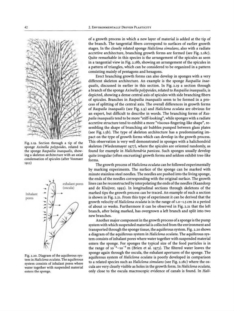
Fig. 2.20.Diagram of the aquiferous system in Haliclona oculata. Theaquiferous
systemconsists of inhalant pores where
water together with suspended material
enters the sponge.
of a growth process in which a new layer of material is added at the tip of
the branch. The tangential fibers correspond to surfaces of earlier growth
stages. In the closely related sponge Haliclona simulans, also with a radiate
accretive architecture, branching growth forms are formed (see Fig.2.18c).
Quite remarkable in this species is the arrangement of the spicules as seen
in a tangential view in Fig. 2.18b, showing an arrangement of the spicules in
a pattern of triangles, which can be considered to be organized in a pattern
consisting mainly of pentagons and hexagons .
Erect branching growth forms can also develop in sponges with a very
different skeleton architecture. An example is the sponge Raspailia inaequalis, discussed in earlier in this section. In Fig. 2.19 a section through
a branch of the sponge Axinella polypoides, related to Raspailia inaequalis, is
depicted, showing a dense central axis of spicules with side branching fibers
of spicules. Branches in Raspailia inaequalis seem to be formed in a pro -
cess of splitting of the central axis. The overall differences in growth forms
of Raspaila inaequalis (see Fig. 2.9) and Haliclona oculata are obvious for
an expert, but difficult to describe in words. The branching forms of Raspaila inaequalis tend to be more "stiff-looking", while sponges with a radiate
accretive structure tend to exhibit a more "viscous-fingering-like shape", resembling the shape of branching air bubbles pumped between glass plates
(see Fig.r.sb). The type of skeleton architecture has a predominating impact on the type of growth forms which can develop in the growth process .
This observation is very well demonstrated in sponges with a halichondrid
skeleton (Wiedenmayer 1977), where the spicules are oriented randomly, as
found for example in Halichondria panicea. Such sponges usually develop
quite irregular (often encrusting) growth forms and seldom exhibit tree-like
forms .
The growth process of Haliclonaoculata can be followed experimentally
by marking experiments. The surface of the sponge can be marked with
minute stainless steel needles. The needles are pushed into the living sponge,
the ends of the needles corresponding with the original surface . The growth
lines can be reconstructed by interpolating the ends of the needles (Kaandorp
and de Kluijver, 1992). In longitudinal sections through skeletons of the
marked tips the growth process can be traced. An example of such a section
is shown in Fig. 2.21. From this type of experiment it can be derived that the
growth velocity of Haliclona oculata is in the range of 1.0-1.5ern in a period
of about 10 weeks. Furthermore it can be observed in Fig. 2.21 that the left
branch, after being marked, has overgrown a left branch and split into two
new branches.
Another major component in the growth process of a sponge is the pump
system with which suspended material is collected from the environment and
transported through the sponge tissue, the aquiferous system. Fig. 2.20 shows
a diagram of the aquiferous system in Haliclonaoculata. The aquiferous system consists of inhalant pores where water together with suspended material
enters the sponge. For sponges the typical size of the food particles is in
the range of 10- 4 -10 -6 m (Brien et al. 1973). The filtered water leaves the
sponge again through the oscula, the exhalant apertures of the sponge. The
aquiferous system of Haliclona oculata is poorly developed in comparison
to a related species such as Haliclona simulans (see Fig. 2.18c) where the oscula are very clearly visible as holes in the growth form . In Haliclonaoculata,
only close to the oscula macroscopic evidence of canals is found. In Haliexhalant pores
(oscula)
Inhalant
pores
~
i
,, ---; r-.!'
% - \' , ~
,.-f I
/7~I
~LiJ<,
-
Fig.2.19. Section through a tip of the
sponge Axinella polypoides, related to
the sponge Raspailia inaequalis, showing a skeleton architecture with an axial
condensation of spicules (after Vosmaer
1912)
2. ENVIRONMENTALLY DRIVEN PLASTICITY
Fig. 2.20.Diagram of the aquiferous system in Haliclona oculata. Theaquiferous
systemconsists of inhalant pores where
water together with suspended material
enters the sponge.
of a growth process in which a new layer of material is added at the tip of
the branch. The tangential fibers correspond to surfaces of earlier growth
stages. In the closely related sponge Haliclona simulans, also with a radiate
accretive architecture, branching growth forms are formed (see Fig.2.18c).
Quite remarkable in this species is the arrangement of the spicules as seen
in a tangential view in Fig. 2.18b, showing an arrangement of the spicules in
a pattern of triangles, which can be considered to be organized in a pattern
consisting mainly of pentagons and hexagons .
Erect branching growth forms can also develop in sponges with a very
different skeleton architecture. An example is the sponge Raspailia inaequalis, discussed in earlier in this section. In Fig. 2.19 a section through
a branch of the sponge Axinella polypoides, related to Raspailia inaequalis, is
depicted, showing a dense central axis of spicules with side branching fibers
of spicules. Branches in Raspailia inaequalis seem to be formed in a pro -
cess of splitting of the central axis. The overall differences in growth forms
of Raspaila inaequalis (see Fig. 2.9) and Haliclona oculata are obvious for
an expert, but difficult to describe in words. The branching forms of Raspaila inaequalis tend to be more "stiff-looking", while sponges with a radiate
accretive structure tend to exhibit a more "viscous-fingering-like shape", resembling the shape of branching air bubbles pumped between glass plates
(see Fig.r.sb). The type of skeleton architecture has a predominating impact on the type of growth forms which can develop in the growth process .
This observation is very well demonstrated in sponges with a halichondrid
skeleton (Wiedenmayer 1977), where the spicules are oriented randomly, as
found for example in Halichondria panicea. Such sponges usually develop
quite irregular (often encrusting) growth forms and seldom exhibit tree-like
forms .
The growth process of Haliclonaoculata can be followed experimentally
by marking experiments. The surface of the sponge can be marked with
minute stainless steel needles. The needles are pushed into the living sponge,
the ends of the needles corresponding with the original surface . The growth
lines can be reconstructed by interpolating the ends of the needles (Kaandorp
and de Kluijver, 1992). In longitudinal sections through skeletons of the
marked tips the growth process can be traced. An example of such a section
is shown in Fig. 2.21. From this type of experiment it can be derived that the
growth velocity of Haliclona oculata is in the range of 1.0-1.5ern in a period
of about 10 weeks. Furthermore it can be observed in Fig. 2.21 that the left
branch, after being marked, has overgrown a left branch and split into two
new branches.
Another major component in the growth process of a sponge is the pump
system with which suspended material is collected from the environment and
transported through the sponge tissue, the aquiferous system. Fig. 2.20 shows
a diagram of the aquiferous system in Haliclonaoculata. The aquiferous system consists of inhalant pores where water together with suspended material
enters the sponge. For sponges the typical size of the food particles is in
the range of 10- 4 -10 -6 m (Brien et al. 1973). The filtered water leaves the
sponge again through the oscula, the exhalant apertures of the sponge. The
aquiferous system of Haliclona oculata is poorly developed in comparison
to a related species such as Haliclona simulans (see Fig. 2.18c) where the oscula are very clearly visible as holes in the growth form . In Haliclonaoculata,
only close to the oscula macroscopic evidence of canals is found. In Haliexhalant pores
(oscula)
Inhalant
pores
~
i
,, ---; r-.!'
% - \' , ~
,.-f I
/7~I
~LiJ<,
-
Fig.2.19. Section through a tip of the
sponge Axinella polypoides, related to
the sponge Raspailia inaequalis, showing a skeleton architecture with an axial
condensation of spicules (after Vosmaer
1912)