

3.4.THREE-DIMENSIONAL MORPHOLOGICAL ANALYSIS OF GROWTH FORMS OF MADRACIS MIRABILIS
3-4 Three-Dimensional Morphological Analysis
of Growth Forms of Madracis Mirabilis
(Preliminary Results)
87
In Sect. 3.3the morphological analysis of two-dimensional images of growth
forms of the three species, collected along environmental gradients, was
discussed in detail. The morphological analysis only partly works using
two-dimensional images; for complex-shaped growth forms with many overlapping branches, as shown in Fig. 3.11, a two-dimensional analysis fails and
a three-dimensional analysis is required. In this section we will present some
preliminary results on how similar methods, as discussed in Sect. 3.3, can be
extended towards a three-dimensional analysis. Since a complete set of measurements is not yet available (will be described in Vermeij et al. in prep.), we
will focus on the three-dimensional data acquisition and give a short description of the thinning procedure, the construction of morphological skeletons,
and the measurements based on these skeletons, for the three-dimensional
case.
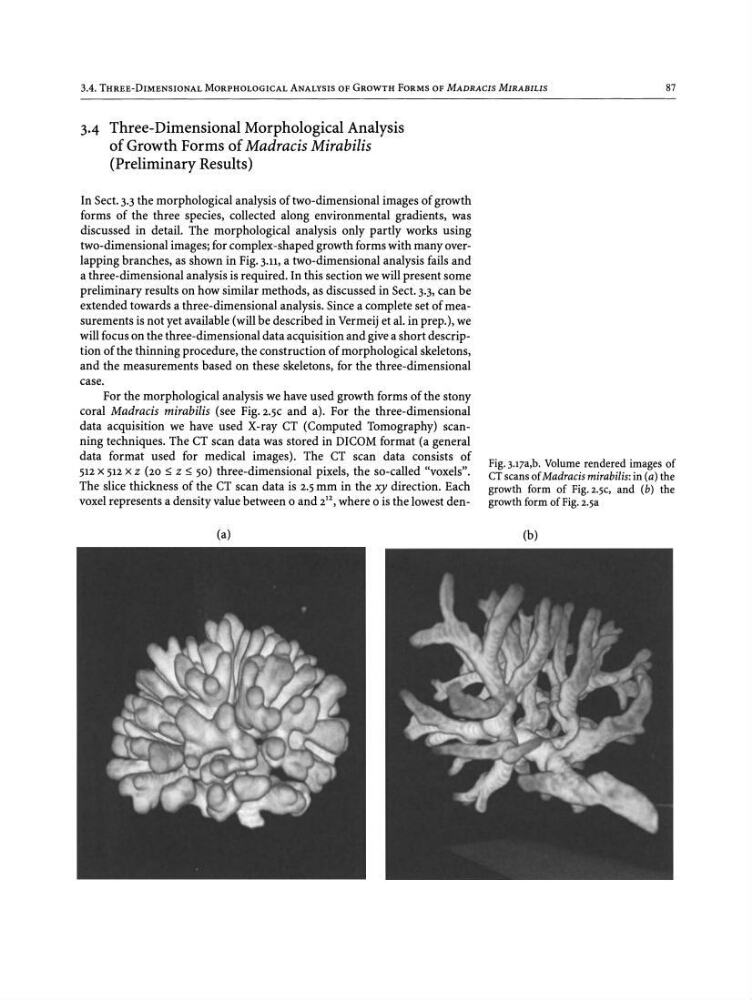
For the morphological analysis we have used growth forms of the stony
coral Madracis mirabilis (see Pig.z.sc and a). For the three-dimensional
data acquisition we have used X-ray CT (Computed Tomography) scanning techniques. The CT scan data was stored in DICOM format (a general
data format used for medical images). The CT scan data consists of
512 x 512 X Z (20 :s; Z :s; 50) three-dimensional pixels, the so-called "voxels",
The slice thickness of the CT scan data is 2.5 mm in the xy direction. Each
voxel represents a density value between 0 and 2
12
, where 0 is the lowest den(a)
Pig.j.iza.b. Volume rendered images of
CT scans of Madracis mirabilis: in (a) the
growth form of Fig.2.5C, and (b) the
growth form of Fig. 2.5a
(b)
3-4 Three-Dimensional Morphological Analysis
of Growth Forms of Madracis Mirabilis
(Preliminary Results)
87
In Sect. 3.3the morphological analysis of two-dimensional images of growth
forms of the three species, collected along environmental gradients, was
discussed in detail. The morphological analysis only partly works using
two-dimensional images; for complex-shaped growth forms with many overlapping branches, as shown in Fig. 3.11, a two-dimensional analysis fails and
a three-dimensional analysis is required. In this section we will present some
preliminary results on how similar methods, as discussed in Sect. 3.3, can be
extended towards a three-dimensional analysis. Since a complete set of measurements is not yet available (will be described in Vermeij et al. in prep.), we
will focus on the three-dimensional data acquisition and give a short description of the thinning procedure, the construction of morphological skeletons,
and the measurements based on these skeletons, for the three-dimensional
case.
For the morphological analysis we have used growth forms of the stony
coral Madracis mirabilis (see Pig.z.sc and a). For the three-dimensional
data acquisition we have used X-ray CT (Computed Tomography) scanning techniques. The CT scan data was stored in DICOM format (a general
data format used for medical images). The CT scan data consists of
512 x 512 X Z (20 :s; Z :s; 50) three-dimensional pixels, the so-called "voxels",
The slice thickness of the CT scan data is 2.5 mm in the xy direction. Each
voxel represents a density value between 0 and 2
12
, where 0 is the lowest den(a)
Pig.j.iza.b. Volume rendered images of
CT scans of Madracis mirabilis: in (a) the
growth form of Fig.2.5C, and (b) the
growth form of Fig. 2.5a
(b)