

68
Methods for Setting Up Primary Cultures Specific to Animal Groups
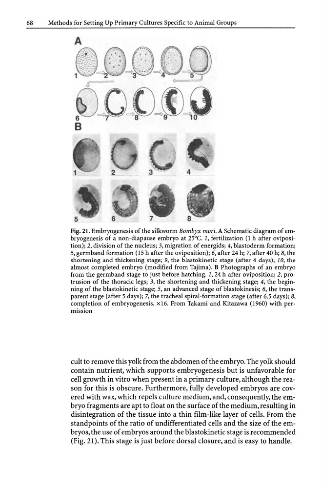
Fig. 21. Embryogenesis of the silkworm Bombyx mori. A Schematic diagram of embryogenesis of a non-diapause embryo at 25°C. 1, fertilization (1 h after oviposition); 2, division of the nucleus; 3, migration of energids; 4, blastoderm formation;
5, germband formation (15 h after the oviposition); 6, after 24 h; 7, after 40 h; 8, the
shortening and thickening stage; 9, the blastokinetic stage (after 4 days); 10, the
almost completed embryo (modified from Tajima). B Photographs of an embryo
from the germband stage to just before hatching. 1,24 h after oviposition; 2, protrusion of the thoracic legs; 3, the shortening and thickening stage; 4, the beginning of the blastokinetic stage; 5, an advanced stage of blastokinesis; 6, the transparent stage (after 5 days); 7, the tracheal spiral-formation stage (after 6.5 days); 8,
completion of embryogenesis. x16. From Takami and Kitazawa (1960) with permission
cult to remove this yolk from the abdomen of the embryo. The yolk should
contain nutrient, which supports embryogenesis but is unfavorable for
cell growth in vitro when present in a primary culture, although the reason for this is obscure. Furthermore, fully developed embryos are covered with wax, which repels culture medium, and, consequently, the embryo fragments are apt to float on the surface of the medium, resulting in
disintegration of the tissue into a thin film-like layer of cells. From the
standpoints of the ratio of undifferentiated cells and the size of the embryos, the use of embryos around the blastokinetic stage is recommended
(Fig. 21). This stage is just before dorsal closure, and is easy to handle.
Methods for Setting Up Primary Cultures Specific to Animal Groups
Fig. 21. Embryogenesis of the silkworm Bombyx mori. A Schematic diagram of embryogenesis of a non-diapause embryo at 25°C. 1, fertilization (1 h after oviposition); 2, division of the nucleus; 3, migration of energids; 4, blastoderm formation;
5, germband formation (15 h after the oviposition); 6, after 24 h; 7, after 40 h; 8, the
shortening and thickening stage; 9, the blastokinetic stage (after 4 days); 10, the
almost completed embryo (modified from Tajima). B Photographs of an embryo
from the germband stage to just before hatching. 1,24 h after oviposition; 2, protrusion of the thoracic legs; 3, the shortening and thickening stage; 4, the beginning of the blastokinetic stage; 5, an advanced stage of blastokinesis; 6, the transparent stage (after 5 days); 7, the tracheal spiral-formation stage (after 6.5 days); 8,
completion of embryogenesis. x16. From Takami and Kitazawa (1960) with permission
cult to remove this yolk from the abdomen of the embryo. The yolk should
contain nutrient, which supports embryogenesis but is unfavorable for
cell growth in vitro when present in a primary culture, although the reason for this is obscure. Furthermore, fully developed embryos are covered with wax, which repels culture medium, and, consequently, the embryo fragments are apt to float on the surface of the medium, resulting in
disintegration of the tissue into a thin film-like layer of cells. From the
standpoints of the ratio of undifferentiated cells and the size of the embryos, the use of embryos around the blastokinetic stage is recommended
(Fig. 21). This stage is just before dorsal closure, and is easy to handle.