

296
Organ Culture
When Sengel (1960) cultured the blastema formed on the transverse
cut surface of Dugesia lugubris 3 days after operation in her culture medium, the explants survived for 10-15 days. During that period, differentiation of pigment cells, muscles, eyes, and brain occurred if the blastema was derived from the head region.
Subprotocol 2
Scolex of Cyclophyllidean Cestodes (Cestoda)
• • Materials
Equipment - Dissecting microscope
- Roller drum and roller tubes
- Scissors
- Forceps
- Needles
- Pasteur pipettes
Solutions - Ringer's solution
- Gey's solution (see Appendix 1)
- Culture medium (50% horse serum-Gey's solution with glucose)
Animals - Adult Hymenolepis diminuta from rodents
• • Procedure
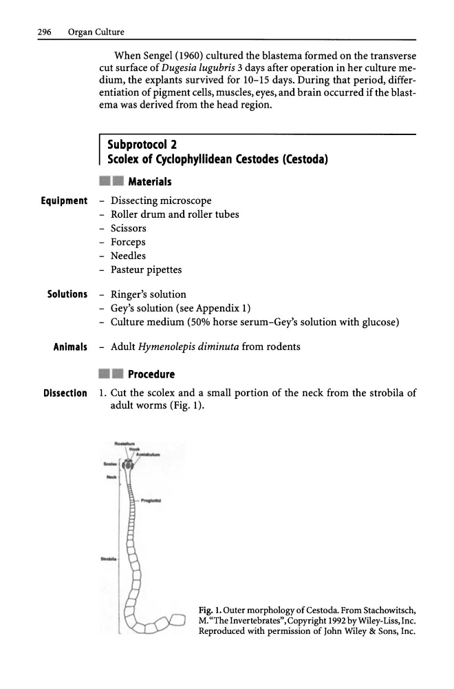
Dissection 1. Cut the scolex and a small portion of the neck from the strobila of
adult worms (Fig. 1).
- - -
- -
-
-
Fig. 1. Outer morphology of Cestoda. From Stachowitsch,
M. "The Invertebrates", Copyright 1992 by Wiley-Liss, Inc.
Reproduced with permission of John Wiley & Sons, Inc.
Organ Culture
When Sengel (1960) cultured the blastema formed on the transverse
cut surface of Dugesia lugubris 3 days after operation in her culture medium, the explants survived for 10-15 days. During that period, differentiation of pigment cells, muscles, eyes, and brain occurred if the blastema was derived from the head region.
Subprotocol 2
Scolex of Cyclophyllidean Cestodes (Cestoda)
• • Materials
Equipment - Dissecting microscope
- Roller drum and roller tubes
- Scissors
- Forceps
- Needles
- Pasteur pipettes
Solutions - Ringer's solution
- Gey's solution (see Appendix 1)
- Culture medium (50% horse serum-Gey's solution with glucose)
Animals - Adult Hymenolepis diminuta from rodents
• • Procedure
Dissection 1. Cut the scolex and a small portion of the neck from the strobila of
adult worms (Fig. 1).
- - -
- -
-
-
Fig. 1. Outer morphology of Cestoda. From Stachowitsch,
M. "The Invertebrates", Copyright 1992 by Wiley-Liss, Inc.
Reproduced with permission of John Wiley & Sons, Inc.