

290
Organ Culture
re
a
de
e
pt
dv
em
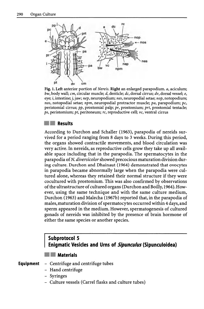
Fig. 1. Left anterior portion of Nereis. Right an enlarged parapodium. a, aciculum;
bw, body wall; em, circular muscle; d, denticle; dc, dorsal cirrus; dv, dorsal vessel; e,
eye; i, intestine; j, jaw; nep, neuropodium; nes, neuropodial setae; nap, notopodium;
nos, notopodial setae; npm, neuropodial protractor muscle; pa, parapodium; pc,
peristomial cirrus; pp, prostomial palp; pr, prostomium; prt, prostomial tentacle;
ps, peristomium; pt, peritoneum; re, reproductive cell; ve, ventral cirrus
• • Results
According to Durchon and Schaller (1963), parapodia of nereids survived for a period ranging from 8 days to 3 weeks. During this period,
the organs showed contractile movements, and blood circulation was
very active. In nereids, as reproductive cells grow they take up all available space including that in the parapodia. The spermatocytes in the
parapodia of N. diversicolor showed precocious maturation division during culture. Durchon and Dhainaut (1964) demonstrated that ovocytes
in parapodia became abnormally large when the parapodia were cultured alone, whereas they retained their normal structure if they were
co cultured with prostomium. This was also confirmed by observations
of the ultrastructure of cultured organs (Durchon and Boilly, 1964). However, using the same technique and with the same culture medium,
Durchon (1963) and Malecha (1967b) reported that, in the parapodia of
males, maturation division of spermatocytes occurred within 4 days, and
sperm appeared in the medium. However, spermatogenesis of cultured
gonads of nereids was inhibited by the presence of brain hormone of
either the same species or another species.
Subprotocol 5
Enigmatic Vesicles and Urns of Sipunculus (Sipunculoidea)
• • Materials
Equipment - Centrifuge and centrifuge tubes
- Hand centrifuge
- Syringes
- Culture vessels (Carrel flasks and culture tubes)
Organ Culture
re
a
de
e
pt
dv
em
Fig. 1. Left anterior portion of Nereis. Right an enlarged parapodium. a, aciculum;
bw, body wall; em, circular muscle; d, denticle; dc, dorsal cirrus; dv, dorsal vessel; e,
eye; i, intestine; j, jaw; nep, neuropodium; nes, neuropodial setae; nap, notopodium;
nos, notopodial setae; npm, neuropodial protractor muscle; pa, parapodium; pc,
peristomial cirrus; pp, prostomial palp; pr, prostomium; prt, prostomial tentacle;
ps, peristomium; pt, peritoneum; re, reproductive cell; ve, ventral cirrus
• • Results
According to Durchon and Schaller (1963), parapodia of nereids survived for a period ranging from 8 days to 3 weeks. During this period,
the organs showed contractile movements, and blood circulation was
very active. In nereids, as reproductive cells grow they take up all available space including that in the parapodia. The spermatocytes in the
parapodia of N. diversicolor showed precocious maturation division during culture. Durchon and Dhainaut (1964) demonstrated that ovocytes
in parapodia became abnormally large when the parapodia were cultured alone, whereas they retained their normal structure if they were
co cultured with prostomium. This was also confirmed by observations
of the ultrastructure of cultured organs (Durchon and Boilly, 1964). However, using the same technique and with the same culture medium,
Durchon (1963) and Malecha (1967b) reported that, in the parapodia of
males, maturation division of spermatocytes occurred within 4 days, and
sperm appeared in the medium. However, spermatogenesis of cultured
gonads of nereids was inhibited by the presence of brain hormone of
either the same species or another species.
Subprotocol 5
Enigmatic Vesicles and Urns of Sipunculus (Sipunculoidea)
• • Materials
Equipment - Centrifuge and centrifuge tubes
- Hand centrifuge
- Syringes
- Culture vessels (Carrel flasks and culture tubes)