

274
Organ Culture
Subprotocol 5
Ventral Eye of Limulus (Merostomacea)
• • Materials
Equipment - Scissors
- Forceps
- Needles
- Pasteur pipettes
- Petri dish
Solutions - Limulus organ culture medium (see Appendix 1)
Animals - Any species of Limulus
• • Procedure
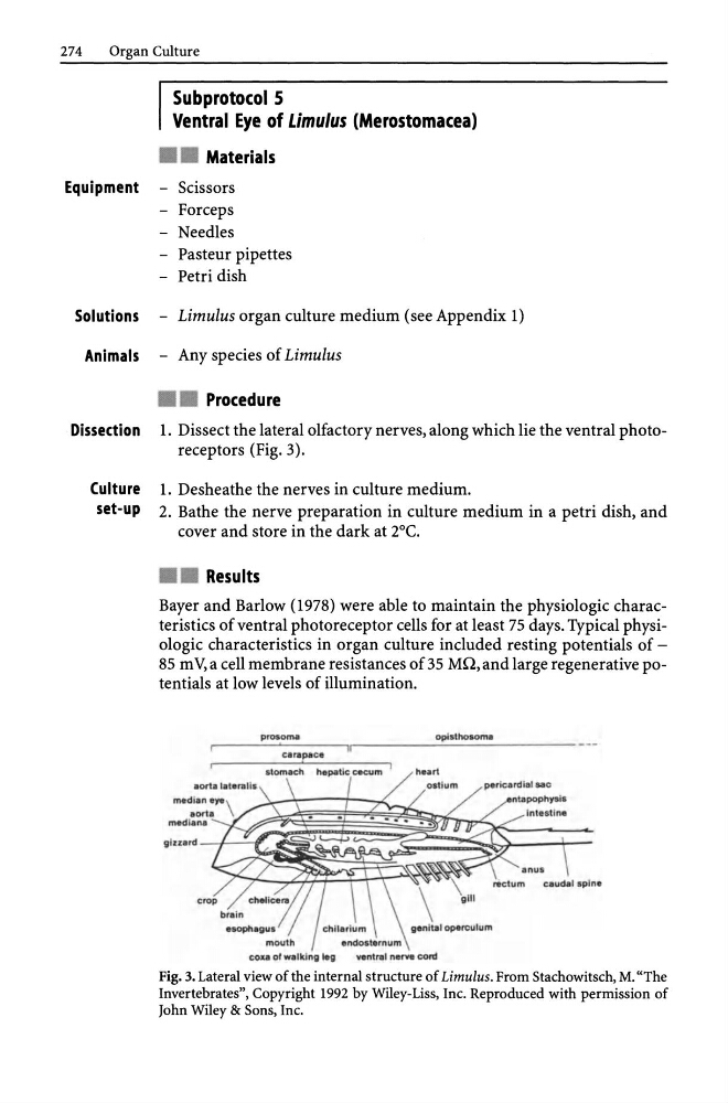
Dissection 1. Dissect the lateral olfactory nerves, along which lie the ventral photoreceptors (Fig. 3).
Culture 1. Desheathe the nerves in culture medium.
set-up 2. Bathe the nerve preparation in culture medium in a petri dish, and
cover and store in the dark at 2°C .
• • Results
Bayer and Barlow (1978) were able to maintain the physiologic characteristics of ventral photoreceptor cells for at least 75 days. Typical physiologic characteristics in organ culture included resting potentials of -
85 m V, a cell membrane resistances of 35 MQ, and large regenerative potentials at low levels of illumination.
median eye \
aorta
med lAna
pro' ,oma
eoo of walking leg
opl sthosoma
Fig. 3. Lateral view of the internal structure of Limulus. From Stachowitsch, M. "The
Invertebrates", Copyright 1992 by Wiley-Liss, Inc. Reproduced with permission of
John Wiley & Sons, Inc.
Organ Culture
Subprotocol 5
Ventral Eye of Limulus (Merostomacea)
• • Materials
Equipment - Scissors
- Forceps
- Needles
- Pasteur pipettes
- Petri dish
Solutions - Limulus organ culture medium (see Appendix 1)
Animals - Any species of Limulus
• • Procedure
Dissection 1. Dissect the lateral olfactory nerves, along which lie the ventral photoreceptors (Fig. 3).
Culture 1. Desheathe the nerves in culture medium.
set-up 2. Bathe the nerve preparation in culture medium in a petri dish, and
cover and store in the dark at 2°C .
• • Results
Bayer and Barlow (1978) were able to maintain the physiologic characteristics of ventral photoreceptor cells for at least 75 days. Typical physiologic characteristics in organ culture included resting potentials of -
85 m V, a cell membrane resistances of 35 MQ, and large regenerative potentials at low levels of illumination.
median eye \
aorta
med lAna
pro' ,oma
eoo of walking leg
opl sthosoma
Fig. 3. Lateral view of the internal structure of Limulus. From Stachowitsch, M. "The
Invertebrates", Copyright 1992 by Wiley-Liss, Inc. Reproduced with permission of
John Wiley & Sons, Inc.