

21 Insecta
247
- Petri dishes
- Culture vessels
- Cover glasses
- Physiological solution
Solutions
- Culture medium (mixture of 5 parts Schneider's insect solution and 4
parts Eagle's medium)
- Nymphs of the American cockroach P. americana (4-6 months old)
Animals
• • Procedure
1. Starve the animals for 1 week.
2. Anesthetize the animals with CO2,
3. Surface sterilize the animals by immersion in 2% iodine and three
changes of 95% ethanol.
4. Wash the animals with sterilized distilled water.
Sterilization
1. Decapitate the animals, and place the cut head in the depression of a Dissection
Maximov slide filled with physiological solution.
2. Quickly excise the brain with the surrounding tissues, and wash it
repeatedly with physiological solution.
3. Separate the frontal ganglion from the tritocerebrum, to which the
frontal ganglion closely adheres, by cutting the two large frontal
connectives, which run from the anterior ends of the ganglion to the
tritocerebrum. Cut the other small nerves connecting the ganglion to
the buccal apparatus and pharynx. Then, also cut the large recurrent
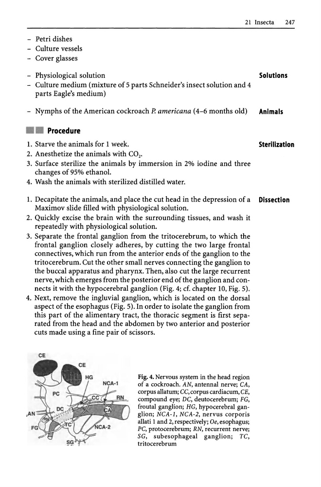
nerve, which emerges from the posterior end of the ganglion and connects it with the hypo cerebral ganglion (Fig. 4; d. chapter 10, Fig. 5).
4. Next, remove the ingluvial ganglion, which is located on the dorsal
aspect of the esophagus (Fig. 5). In order to isolate the ganglion from
this part of the alimentary tract, the thoracic segment is first separated from the head and the abdomen by two anterior and posterior
cuts made using a fine pair of scissors.
CE
Fig. 4. Nervous system in the head region
of a cockroach. AN, antennal nerve; CA,
corpus allatum; CC, corpus cardiacum, CE,
compound eye; DC, deutocerebrum; FG,
froutal ganglion; HG, hypocerebral ganglion; NCA-l, NCA-2, nervus corporis
allati 1 and 2, respectively; Oe, esophagus;
PC, proto cerebrum; RN, recurrent nerve;
SG, subesophageal ganglion; TC,
tritocerebrum
247
- Petri dishes
- Culture vessels
- Cover glasses
- Physiological solution
Solutions
- Culture medium (mixture of 5 parts Schneider's insect solution and 4
parts Eagle's medium)
- Nymphs of the American cockroach P. americana (4-6 months old)
Animals
• • Procedure
1. Starve the animals for 1 week.
2. Anesthetize the animals with CO2,
3. Surface sterilize the animals by immersion in 2% iodine and three
changes of 95% ethanol.
4. Wash the animals with sterilized distilled water.
Sterilization
1. Decapitate the animals, and place the cut head in the depression of a Dissection
Maximov slide filled with physiological solution.
2. Quickly excise the brain with the surrounding tissues, and wash it
repeatedly with physiological solution.
3. Separate the frontal ganglion from the tritocerebrum, to which the
frontal ganglion closely adheres, by cutting the two large frontal
connectives, which run from the anterior ends of the ganglion to the
tritocerebrum. Cut the other small nerves connecting the ganglion to
the buccal apparatus and pharynx. Then, also cut the large recurrent
nerve, which emerges from the posterior end of the ganglion and connects it with the hypo cerebral ganglion (Fig. 4; d. chapter 10, Fig. 5).
4. Next, remove the ingluvial ganglion, which is located on the dorsal
aspect of the esophagus (Fig. 5). In order to isolate the ganglion from
this part of the alimentary tract, the thoracic segment is first separated from the head and the abdomen by two anterior and posterior
cuts made using a fine pair of scissors.
CE
Fig. 4. Nervous system in the head region
of a cockroach. AN, antennal nerve; CA,
corpus allatum; CC, corpus cardiacum, CE,
compound eye; DC, deutocerebrum; FG,
froutal ganglion; HG, hypocerebral ganglion; NCA-l, NCA-2, nervus corporis
allati 1 and 2, respectively; Oe, esophagus;
PC, proto cerebrum; RN, recurrent nerve;
SG, subesophageal ganglion; TC,
tritocerebrum