

210
Methods for Setting Up Primary Cultures Specific to Animal Groups
Dissociation 1. Transfer the cysts into a petri dish, and mince using scissors.
2. Digest the minced cysts with 0.25% trypsin solution at ambient temperature for 30 min.
3. Filter the digested tissues through a stainless steel mesh (no. 200).
4. Wash the separated cells twice with RPMI-1640 medium by centrifugation at 500 g for 5 min.
Culture 1. Suspend the cell pellet in RPMI-I640 medium supplemented with
set-up
penicillin (500 IU/ml), streptomycin (500 Ilg/ml), 2 mM L-glutamine,
1 roM sodium pyruvate, and 10% FBS.
2. Distribute 2 ml of this cell suspension to collagen-coated plastic dishes.
3. Keep the culture in a 5% CO 2 incubator.
4. Replace the culture medium every 2 or 3 days .
• • Results
Benex (1968) cultured scolex from Echinococcus granulosus from hydatid cysts of a horse in hydatid fluid or synthetic medium, such as TC199, and noticed that some fragments of the germinal membrane were
necessary to obtain true miniature hydatid cysts with a laminated enve02mm
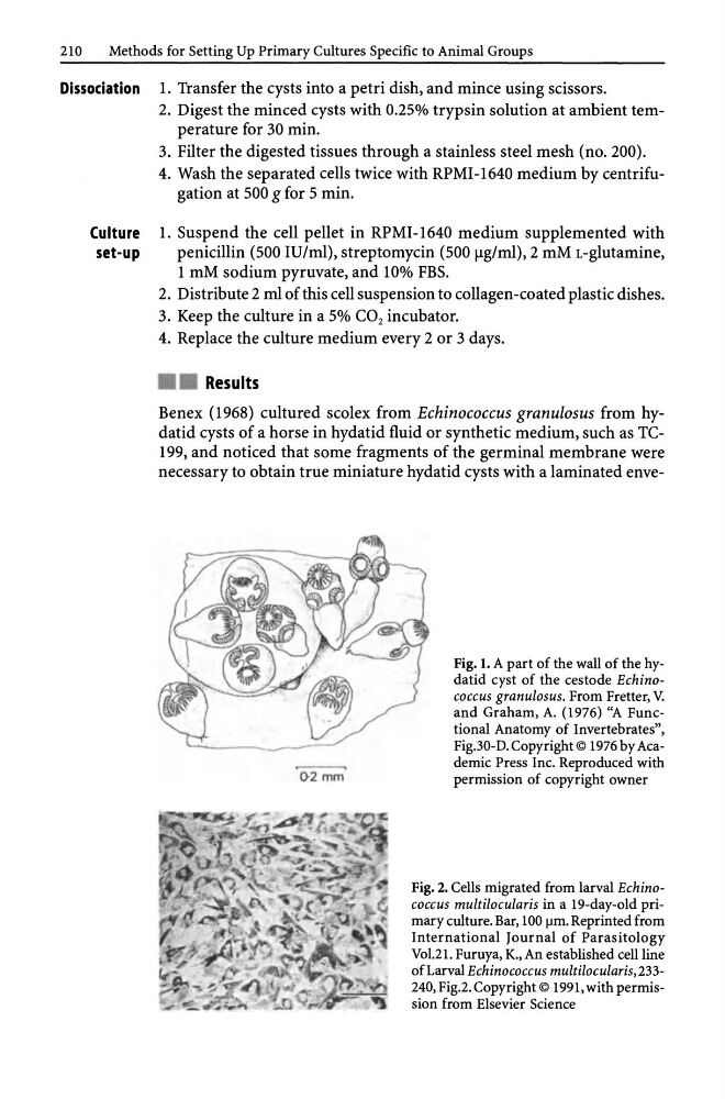
Fig. 1. A part of the wall of the hydatid cyst of the cestode Echinococcus granulosus. From Fretter, V.
and Graham, A. (1976) "A Functional Anatomy of Invertebrates",
Fig.30-D. Copyright © 1976 by Academic Press Inc. Reproduced with
permission of copyright owner
Fig. 2. Cells migrated from larval Echinococcus multilocularis in a 19-day-old primary culture. Bar, 100 f.lm. Reprinted from
International Journal of Parasitology
Vo1.21. Furuya, K., An established cell line
of Larval Echinococcus multilocularis, 233240, Fig.2. Copyright © 1991, with permission from Elsevier Science
Methods for Setting Up Primary Cultures Specific to Animal Groups
Dissociation 1. Transfer the cysts into a petri dish, and mince using scissors.
2. Digest the minced cysts with 0.25% trypsin solution at ambient temperature for 30 min.
3. Filter the digested tissues through a stainless steel mesh (no. 200).
4. Wash the separated cells twice with RPMI-1640 medium by centrifugation at 500 g for 5 min.
Culture 1. Suspend the cell pellet in RPMI-I640 medium supplemented with
set-up
penicillin (500 IU/ml), streptomycin (500 Ilg/ml), 2 mM L-glutamine,
1 roM sodium pyruvate, and 10% FBS.
2. Distribute 2 ml of this cell suspension to collagen-coated plastic dishes.
3. Keep the culture in a 5% CO 2 incubator.
4. Replace the culture medium every 2 or 3 days .
• • Results
Benex (1968) cultured scolex from Echinococcus granulosus from hydatid cysts of a horse in hydatid fluid or synthetic medium, such as TC199, and noticed that some fragments of the germinal membrane were
necessary to obtain true miniature hydatid cysts with a laminated enve02mm
Fig. 1. A part of the wall of the hydatid cyst of the cestode Echinococcus granulosus. From Fretter, V.
and Graham, A. (1976) "A Functional Anatomy of Invertebrates",
Fig.30-D. Copyright © 1976 by Academic Press Inc. Reproduced with
permission of copyright owner
Fig. 2. Cells migrated from larval Echinococcus multilocularis in a 19-day-old primary culture. Bar, 100 f.lm. Reprinted from
International Journal of Parasitology
Vo1.21. Furuya, K., An established cell line
of Larval Echinococcus multilocularis, 233240, Fig.2. Copyright © 1991, with permission from Elsevier Science