

170
Methods for Setting Up Primary Cultures Specific to Animal Groups
- Dispase solution (1,000 U/ml dispase II solution in MMBSS containing 10% pearl oyster hemolymph)
Animals - Pearl oyster P. fucata martensii with a 50-80 mm shell height
• • Procedure
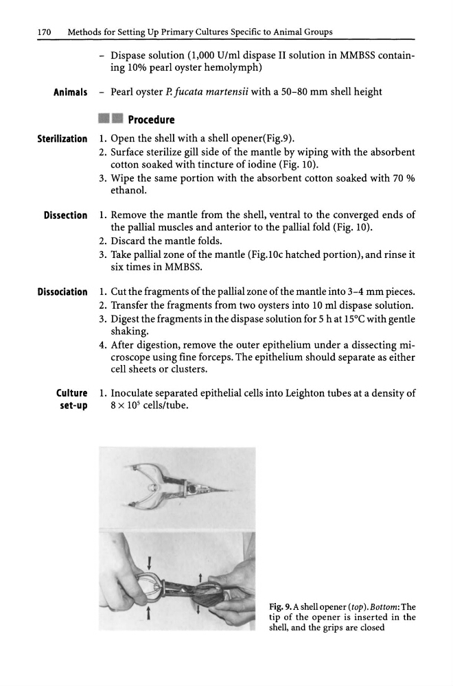
Sterilization 1. Open the shell with a shell opener(Fig.9).
2. Surface sterilize gill side of the mantle by wiping with the absorbent
cotton soaked with tincture of iodine (Fig. 10).
3. Wipe the same portion with the absorbent cotton soaked with 70 %
ethanol.
Dissection 1. Remove the mantle from the shell, ventral to the converged ends of
the pallial muscles and anterior to the pallial fold (Fig. 10).
2. Discard the mantle folds.
3. Take pallial zone of the mantle (Fig.IOc hatched portion), and rinse it
six times in MMBSS.
Dissociation 1. Cut the fragments of the pallial zone of the mantle into 3-4 mm pieces.
2. Transfer the fragments from two oysters into 10 ml dispase solution.
3. Digest the fragments in the dispase solution for 5 h at 15°C with gentle
shaking.
4. After digestion, remove the outer epithelium under a dissecting microscope using fine forceps. The epithelium should separate as either
cell sheets or clusters.
Culture 1. Inoculate separated epithelial cells into Leighton tubes at a density of
set-up
8 x 10 5 cells/tube.
Fig. 9.A shell opener (top ).Bottom: The
tip of the opener is inserted in the
shell, and the grips are closed
Methods for Setting Up Primary Cultures Specific to Animal Groups
- Dispase solution (1,000 U/ml dispase II solution in MMBSS containing 10% pearl oyster hemolymph)
Animals - Pearl oyster P. fucata martensii with a 50-80 mm shell height
• • Procedure
Sterilization 1. Open the shell with a shell opener(Fig.9).
2. Surface sterilize gill side of the mantle by wiping with the absorbent
cotton soaked with tincture of iodine (Fig. 10).
3. Wipe the same portion with the absorbent cotton soaked with 70 %
ethanol.
Dissection 1. Remove the mantle from the shell, ventral to the converged ends of
the pallial muscles and anterior to the pallial fold (Fig. 10).
2. Discard the mantle folds.
3. Take pallial zone of the mantle (Fig.IOc hatched portion), and rinse it
six times in MMBSS.
Dissociation 1. Cut the fragments of the pallial zone of the mantle into 3-4 mm pieces.
2. Transfer the fragments from two oysters into 10 ml dispase solution.
3. Digest the fragments in the dispase solution for 5 h at 15°C with gentle
shaking.
4. After digestion, remove the outer epithelium under a dissecting microscope using fine forceps. The epithelium should separate as either
cell sheets or clusters.
Culture 1. Inoculate separated epithelial cells into Leighton tubes at a density of
set-up
8 x 10 5 cells/tube.
Fig. 9.A shell opener (top ).Bottom: The
tip of the opener is inserted in the
shell, and the grips are closed