

138
Methods for Setting Up Primary Cultures Specific to Animal Groups
• • Procedure
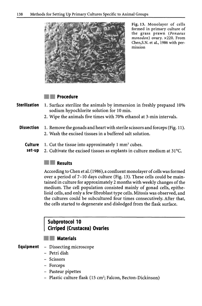
Fig. 13. Monolayer of cells
formed in primary culture of
the grass prawn (Penaeus
monodon) ovary. x220. From
Chen,S.N. et ai., 1986 with permission
Sterilization 1. Surface sterilize the animals by immersion in freshly prepared 10%
sodium hypochlorite solution for 10 min.
2. Wipe the animals five times with 70% ethanol at 3-min intervals.
Dissection 1. Remove the gonads and heart with sterile scissors and forceps (Fig. 11).
2. Wash the excised tissues in a buffered salt solution.
Culture 1. Cut the tissue into approximately 1 mm 3 cubes.
set-up 2. Cultivate the excised tissues as explants in culture medium at 31°C .
• • Results
According to Chen et al. (1986), a confluent monolayer of cells was formed
over a period of 7-10 days culture (Fig. 13). These cells could be maintained in culture for approximately 2 months with weekly changes of the
medium. The cell population consisted mainly of gonad cells, epithelioid cells, and only a few fibroblast type cells. Mitosis was observed, and
the cultures could be subcultured four times consecutively. After that,
the cells started to degenerate and dislodged from the flask surface.
Subprotocol 10
Cirriped (Crustacea) Ovaries
• • Materials
Equipment - Dissecting microscope
- Petri dish
- Scissors
- Forceps
- Pasteur pipettes
- Plastic culture flask (15 cm 2 ; Falcon, Becton-Dickinson)
Methods for Setting Up Primary Cultures Specific to Animal Groups
• • Procedure
Fig. 13. Monolayer of cells
formed in primary culture of
the grass prawn (Penaeus
monodon) ovary. x220. From
Chen,S.N. et ai., 1986 with permission
Sterilization 1. Surface sterilize the animals by immersion in freshly prepared 10%
sodium hypochlorite solution for 10 min.
2. Wipe the animals five times with 70% ethanol at 3-min intervals.
Dissection 1. Remove the gonads and heart with sterile scissors and forceps (Fig. 11).
2. Wash the excised tissues in a buffered salt solution.
Culture 1. Cut the tissue into approximately 1 mm 3 cubes.
set-up 2. Cultivate the excised tissues as explants in culture medium at 31°C .
• • Results
According to Chen et al. (1986), a confluent monolayer of cells was formed
over a period of 7-10 days culture (Fig. 13). These cells could be maintained in culture for approximately 2 months with weekly changes of the
medium. The cell population consisted mainly of gonad cells, epithelioid cells, and only a few fibroblast type cells. Mitosis was observed, and
the cultures could be subcultured four times consecutively. After that,
the cells started to degenerate and dislodged from the flask surface.
Subprotocol 10
Cirriped (Crustacea) Ovaries
• • Materials
Equipment - Dissecting microscope
- Petri dish
- Scissors
- Forceps
- Pasteur pipettes
- Plastic culture flask (15 cm 2 ; Falcon, Becton-Dickinson)