

134
Methods for Setting Up Primary Cultures Specific to Animal Groups
5
• • Results
Fig. 8. The structure of a shrimp leg. 1,
coxopodite; 2, basiopodite; 3, exopodite; 4,
ischiopodite; 5, mesopodite; 6, carpopodite; 7,
propodus; 8, dactylopodite
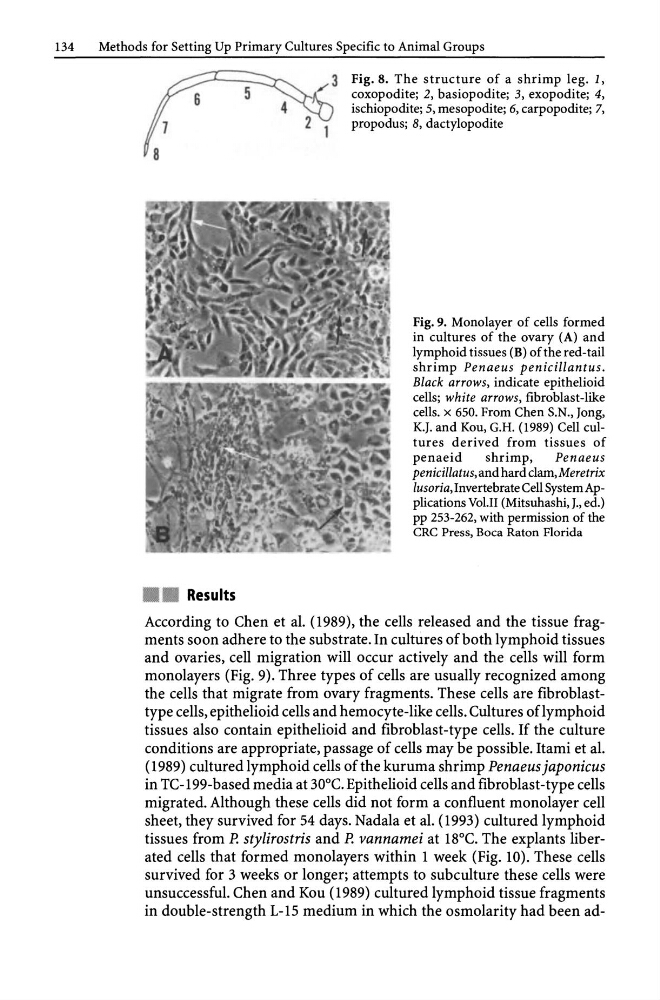
Fig. 9. Monolayer of cells formed
in cultures of the ovary (A) and
lymphoid tissues (B) of the red-tail
shrimp Penaeus penicillantus.
Black arrows, indicate epithelioid
cells; white arrows, fibroblast-like
cells. x 650. From Chen S.N., Jong,
K.J. and Kou, G.H. (1989) Cell cultures derived from tissues of
penaeid
shrimp,
Penaeus
penicillatus, and hard c1am,Meretrix
lusoria, Invertebrate Cell System Applications Vol.II (Mitsuhashi, J., ed.)
pp 253-262, with permission of the
CRC Press, Boca Raton Florida
According to Chen et al. (1989), the cells released and the tissue fragments soon adhere to the substrate. In cultures of both lymphoid tissues
and ovaries, cell migration will occur actively and the cells will form
monolayers (Fig. 9). Three types of cells are usually recognized among
the cells that migrate from ovary fragments. These cells are fibroblasttype cells, epithelioid cells and hemocyte-like cells. Cultures oflymphoid
tissues also contain epithelioid and fibroblast-type cells. If the culture
conditions are appropriate, passage of cells may be possible. Itami et al.
(1989) cultured lymphoid cells of the kuruma shrimp Penaeus japonicus
in TC-199-based media at 30°C. Epithelioid cells and fibroblast -type cells
migrated. Although these cells did not form a confluent monolayer cell
sheet, they survived for 54 days. Nadala et al. (1993) cultured lymphoid
tissues from P. stylirostris and P. vannamei at 18°C. The explants liberated cells that formed monolayers within 1 week (Fig. 10). These cells
survived for 3 weeks or longer; attempts to subculture these cells were
unsuccessful. Chen and Kou (1989) cultured lymphoid tissue fragments
in double-strength L-15 medium in which the osmolarity had been ad-
Methods for Setting Up Primary Cultures Specific to Animal Groups
5
• • Results
Fig. 8. The structure of a shrimp leg. 1,
coxopodite; 2, basiopodite; 3, exopodite; 4,
ischiopodite; 5, mesopodite; 6, carpopodite; 7,
propodus; 8, dactylopodite
Fig. 9. Monolayer of cells formed
in cultures of the ovary (A) and
lymphoid tissues (B) of the red-tail
shrimp Penaeus penicillantus.
Black arrows, indicate epithelioid
cells; white arrows, fibroblast-like
cells. x 650. From Chen S.N., Jong,
K.J. and Kou, G.H. (1989) Cell cultures derived from tissues of
penaeid
shrimp,
Penaeus
penicillatus, and hard c1am,Meretrix
lusoria, Invertebrate Cell System Applications Vol.II (Mitsuhashi, J., ed.)
pp 253-262, with permission of the
CRC Press, Boca Raton Florida
According to Chen et al. (1989), the cells released and the tissue fragments soon adhere to the substrate. In cultures of both lymphoid tissues
and ovaries, cell migration will occur actively and the cells will form
monolayers (Fig. 9). Three types of cells are usually recognized among
the cells that migrate from ovary fragments. These cells are fibroblasttype cells, epithelioid cells and hemocyte-like cells. Cultures oflymphoid
tissues also contain epithelioid and fibroblast-type cells. If the culture
conditions are appropriate, passage of cells may be possible. Itami et al.
(1989) cultured lymphoid cells of the kuruma shrimp Penaeus japonicus
in TC-199-based media at 30°C. Epithelioid cells and fibroblast -type cells
migrated. Although these cells did not form a confluent monolayer cell
sheet, they survived for 54 days. Nadala et al. (1993) cultured lymphoid
tissues from P. stylirostris and P. vannamei at 18°C. The explants liberated cells that formed monolayers within 1 week (Fig. 10). These cells
survived for 3 weeks or longer; attempts to subculture these cells were
unsuccessful. Chen and Kou (1989) cultured lymphoid tissue fragments
in double-strength L-15 medium in which the osmolarity had been ad-