

126
Methods for Setting Up Primary Cultures Specific to Animal Groups
Central ganglion
Rectal J+--b'IiIfU:;;
sac
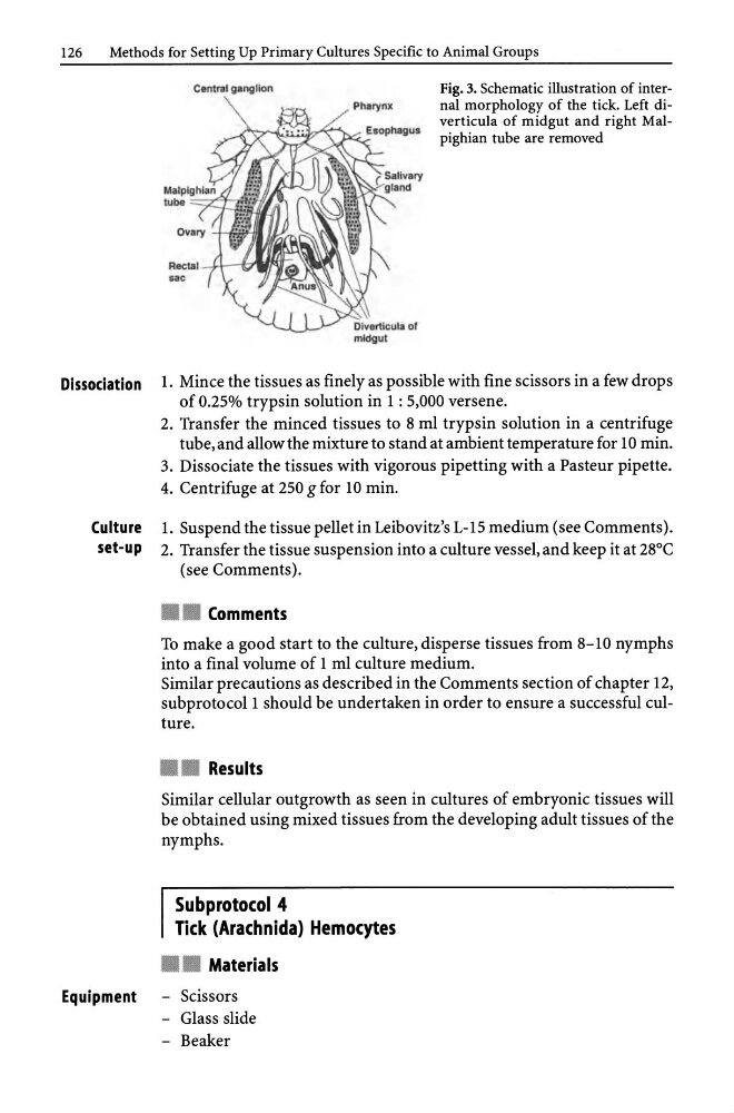
Fig. 3. Schematic illustration of internal morphology of the tick. Left diverticula of midgut and right Malpighian tube are removed
Dissociation 1. Mince the tissues as finely as possible with fine scissors in a few drops
of 0.25% trypsin solution in 1 : 5,000 versene.
2. Transfer the minced tissues to 8 ml trypsin solution in a centrifuge
tube, and allow the mixture to stand at ambient temperature for 10 min.
3. Dissociate the tissues with vigorous pipetting with a Pasteur pipette.
4. Centrifuge at 250 g for 10 min.
Culture l. Suspend the tissue pellet in Leibovitz's L-15 medium (see Comments).
set-up 2. Transfer the tissue suspension into a culture vessel, and keep it at 28°C
(see Comments) .
• • Comments
To make a good start to the culture, disperse tissues from 8-10 nymphs
into a final volume of 1 ml culture medium.
Similar precautions as described in the Comments section of chapter 12,
subprotocoll should be undertaken in order to ensure a successful culture .
• • Results
Similar cellular outgrowth as seen in cultures of embryonic tissues will
be obtained using mixed tissues from the developing adult tissues of the
nymphs.
Subprotocol 4
Tick (Arachnida) Hemocytes
• • Materials
Equipment - Scissors
- Glass slide
- Beaker
Methods for Setting Up Primary Cultures Specific to Animal Groups
Central ganglion
Rectal J+--b'IiIfU:;;
sac
Fig. 3. Schematic illustration of internal morphology of the tick. Left diverticula of midgut and right Malpighian tube are removed
Dissociation 1. Mince the tissues as finely as possible with fine scissors in a few drops
of 0.25% trypsin solution in 1 : 5,000 versene.
2. Transfer the minced tissues to 8 ml trypsin solution in a centrifuge
tube, and allow the mixture to stand at ambient temperature for 10 min.
3. Dissociate the tissues with vigorous pipetting with a Pasteur pipette.
4. Centrifuge at 250 g for 10 min.
Culture l. Suspend the tissue pellet in Leibovitz's L-15 medium (see Comments).
set-up 2. Transfer the tissue suspension into a culture vessel, and keep it at 28°C
(see Comments) .
• • Comments
To make a good start to the culture, disperse tissues from 8-10 nymphs
into a final volume of 1 ml culture medium.
Similar precautions as described in the Comments section of chapter 12,
subprotocoll should be undertaken in order to ensure a successful culture .
• • Results
Similar cellular outgrowth as seen in cultures of embryonic tissues will
be obtained using mixed tissues from the developing adult tissues of the
nymphs.
Subprotocol 4
Tick (Arachnida) Hemocytes
• • Materials
Equipment - Scissors
- Glass slide
- Beaker