

120
Methods for Setting Up Primary Cultures Specific to Animal Groups
• • Results
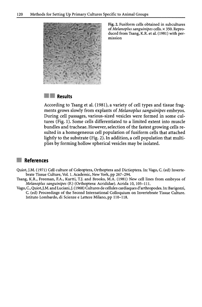
Fig. 2. Fusiform cells obtained in subcultures
of Melanoplus sanguinipes cells. x 350. Reproduced from Tsang, K.R. et al. (1981) with permission
According to Tsang et al. (1981), a variety of cell types and tissue fragments grows slowly from explants of Melanoplus sanguinipes embryos.
During cell passages, various-sized vesicles were formed in some cultures (Fig. 1). Some cells differentiated to a limited extent into muscle
bundles and tracheae. However, selection of the fastest growing cells resulted in a homogeneous cell population of fusiform cells that attached
lightly to the substrate (Fig. 2). In addition, a cell population that multiplies by forming hollow spherical vesicles may be isolated.
• References
Quiot, J.M. (1971) Cell culture of Coleoptera, Orthoptera and Dictioptera. In: Vago, C. (ed) Invertebrate Tissue Culture, Vol. 1. Academic, New York, pp 267-294.
Tsang, K.R., Freeman, F.A., Kurtti, T.J. and Brooks, M.A. (1981) New cell lines from embryos of
Melanoplus sanguinipes (F.) (Orthoptera: Acrididae). Acrida 10, 105-111.
Vago, c., Quiot, J.M. and Luciani, J. (1968) Cultures de cellules cardiaques d' arthropodes. In: Barigozzi,
C. (ed) Proceedings of the Second International Colloquium on Invertebrate Tissue Culture.
Istituto Lombardo, di Scienze e Lettere Milano, pp 11 0-118.
Methods for Setting Up Primary Cultures Specific to Animal Groups
• • Results
Fig. 2. Fusiform cells obtained in subcultures
of Melanoplus sanguinipes cells. x 350. Reproduced from Tsang, K.R. et al. (1981) with permission
According to Tsang et al. (1981), a variety of cell types and tissue fragments grows slowly from explants of Melanoplus sanguinipes embryos.
During cell passages, various-sized vesicles were formed in some cultures (Fig. 1). Some cells differentiated to a limited extent into muscle
bundles and tracheae. However, selection of the fastest growing cells resulted in a homogeneous cell population of fusiform cells that attached
lightly to the substrate (Fig. 2). In addition, a cell population that multiplies by forming hollow spherical vesicles may be isolated.
• References
Quiot, J.M. (1971) Cell culture of Coleoptera, Orthoptera and Dictioptera. In: Vago, C. (ed) Invertebrate Tissue Culture, Vol. 1. Academic, New York, pp 267-294.
Tsang, K.R., Freeman, F.A., Kurtti, T.J. and Brooks, M.A. (1981) New cell lines from embryos of
Melanoplus sanguinipes (F.) (Orthoptera: Acrididae). Acrida 10, 105-111.
Vago, c., Quiot, J.M. and Luciani, J. (1968) Cultures de cellules cardiaques d' arthropodes. In: Barigozzi,
C. (ed) Proceedings of the Second International Colloquium on Invertebrate Tissue Culture.
Istituto Lombardo, di Scienze e Lettere Milano, pp 11 0-118.