

11 Insecta (Orthoptera)
119
2. Dechorionate the eggs by submersion in a 2% aqueous solution of
sodium hypochlorite for 7 min.
3. Rinse the eggs twice with distilled water.
4. Surface sterilize the eggs in a 0.2% aqueous solution of Hyamine 1622
(Rohm and Haas) for 5 min.
5. Transfer the eggs into serum-free culture medium.
1. Dissect the eggs, and remove embryonic tissues with forceps.
Dissection
2. Accumulate embryonic tissues in culture medium placed in a small
petri dish. Tissues from approximately 50 matured embryos or 100150 embryos before or around blastkinetic stage will be needed.
3. Remove as much yolk as possible.
1. Dissociate the tissues by treatment with 0.1 % trypsin in a Ca2+ - and Trypsinization
Mg2+ -free salt solution.
2. Keep the tissues in 2 ml trypsin solution for 4 min with agitation by
pipetting.
3. Stop trypsin digestion by the addition of 5 ml culture medium containing 16.7% FBS.
4. Centrifuge the mixture at 275 g for 5 min.
1. Dispense the cell pellet into 2 ml culture medium with 100 Ilg/ml peni- Culture
cillin G sodium salt and 100 Ilg/ml streptomycin sulfate.
set-up
2. Centrifuge again at 275 g for 5 min.
3. Resuspend the cells in 2 ml culture medium, and dispense into T-15
glass flasks. A seeding density of 10 6 -10 7 cells/ml is suitable.
4. Keep the culture at 26°C.
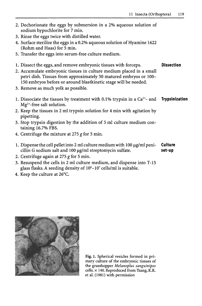
Fig. 1. Spherical vesicles formed in primary culture of the embryonic tissues of
the grasshopper Melanoplus sanguinipes
cells. x 140. Reproduced from Tsang, K.R.
et al. (1981) with permission
119
2. Dechorionate the eggs by submersion in a 2% aqueous solution of
sodium hypochlorite for 7 min.
3. Rinse the eggs twice with distilled water.
4. Surface sterilize the eggs in a 0.2% aqueous solution of Hyamine 1622
(Rohm and Haas) for 5 min.
5. Transfer the eggs into serum-free culture medium.
1. Dissect the eggs, and remove embryonic tissues with forceps.
Dissection
2. Accumulate embryonic tissues in culture medium placed in a small
petri dish. Tissues from approximately 50 matured embryos or 100150 embryos before or around blastkinetic stage will be needed.
3. Remove as much yolk as possible.
1. Dissociate the tissues by treatment with 0.1 % trypsin in a Ca2+ - and Trypsinization
Mg2+ -free salt solution.
2. Keep the tissues in 2 ml trypsin solution for 4 min with agitation by
pipetting.
3. Stop trypsin digestion by the addition of 5 ml culture medium containing 16.7% FBS.
4. Centrifuge the mixture at 275 g for 5 min.
1. Dispense the cell pellet into 2 ml culture medium with 100 Ilg/ml peni- Culture
cillin G sodium salt and 100 Ilg/ml streptomycin sulfate.
set-up
2. Centrifuge again at 275 g for 5 min.
3. Resuspend the cells in 2 ml culture medium, and dispense into T-15
glass flasks. A seeding density of 10 6 -10 7 cells/ml is suitable.
4. Keep the culture at 26°C.
Fig. 1. Spherical vesicles formed in primary culture of the embryonic tissues of
the grasshopper Melanoplus sanguinipes
cells. x 140. Reproduced from Tsang, K.R.
et al. (1981) with permission