

170
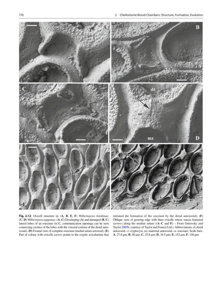
Fig. 2.12 Ovicell structure in: ( A , B , E , F ) Wilbertopora listokinae ;
( C , D ) Wilbertopora tappanae . ( A – C ) Developing ( A ) and damaged ( B , C )
lateral lobes of an ooecium (in C , communication openings can be seen
connecting cavities of the lobes with the visceral coelom of the distal autozooid). ( D ) Frontal view of complete ooecium (medial suture arrowed). ( E )
Part of colony with ovicells ( arrow points to the cryptic avicularium that
initiated the formation of the ooecium by the distal autozooid). ( F )
Oblique view of growing edge with three ovicells whose ooecia fractured
( arrow ) along the median suture (( A – C and F ) – From Ostrovsky and
Taylor 2005b , courtesy of Taylor and Francis Ltd.). Abbreviations: dz distal
autozooid, cr cryptocyst, mz maternal autozooid, oe ooecium. Scale bars:
A , 27.8 μm; B , 40 μm; C , 25.6 μm; D , 34.5 μm; E , 152 μm; F , 156 μm
2 Cheilostome Brood Chambers: Structure, Formation, Evolution
Fig. 2.12 Ovicell structure in: ( A , B , E , F ) Wilbertopora listokinae ;
( C , D ) Wilbertopora tappanae . ( A – C ) Developing ( A ) and damaged ( B , C )
lateral lobes of an ooecium (in C , communication openings can be seen
connecting cavities of the lobes with the visceral coelom of the distal autozooid). ( D ) Frontal view of complete ooecium (medial suture arrowed). ( E )
Part of colony with ovicells ( arrow points to the cryptic avicularium that
initiated the formation of the ooecium by the distal autozooid). ( F )
Oblique view of growing edge with three ovicells whose ooecia fractured
( arrow ) along the median suture (( A – C and F ) – From Ostrovsky and
Taylor 2005b , courtesy of Taylor and Francis Ltd.). Abbreviations: dz distal
autozooid, cr cryptocyst, mz maternal autozooid, oe ooecium. Scale bars:
A , 27.8 μm; B , 40 μm; C , 25.6 μm; D , 34.5 μm; E , 152 μm; F , 156 μm
2 Cheilostome Brood Chambers: Structure, Formation, Evolution