

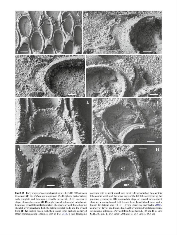
Fig. 2.11 Early stages of ooecium formation in: ( A – E , H ) Wilbertopora
listokinae ; ( F , G ), Wilbertopora tappanae . ( A ) Peripheral part of colony
with complete and developing ovicells ( arrowed ); ( B – H ) successive
stages of ovicellogenesis: ( B – D ) single ooecial rudiment of initial calcifi cation of ovicell fl oor; ( E ) formation of concave ovicell fl oor, showing
skeletal layer underlying both the lateral zooidal walls and the ovicell
fl oor. ( F , G ) Broken ooecia with their lateral lobes partially destroyed
(their communication openings seen in Fig. 2.12C ); ( G ) developing
ooecium with its right lateral lobe mostly detached (short base of this
lobe can be seen), and the lower edge of the left lobe overgrowing the
proximal gymnocyst; ( H ) intermediate stage of ooecial development
showing a hemispherical fold formed from fused lateral lobes and a
broken left lateral lobe (( B – H ) – From Ostrovsky and Taylor 2005b ,
courtesy of Taylor and Francis Ltd.). Abbreviations: dz distal autozooid,
mz maternal autozooid, of ovicell fl oor. Scale bars: A , 125 μm; B , 27 μm;
C , D , 30.3 μm; E , 24.4 μm; F , 20.8 μm; G , 29.4 μm; H , 35.7 μm
listokinae ; ( F , G ), Wilbertopora tappanae . ( A ) Peripheral part of colony
with complete and developing ovicells ( arrowed ); ( B – H ) successive
stages of ovicellogenesis: ( B – D ) single ooecial rudiment of initial calcifi cation of ovicell fl oor; ( E ) formation of concave ovicell fl oor, showing
skeletal layer underlying both the lateral zooidal walls and the ovicell
fl oor. ( F , G ) Broken ooecia with their lateral lobes partially destroyed
(their communication openings seen in Fig. 2.12C ); ( G ) developing
ooecium with its right lateral lobe mostly detached (short base of this
lobe can be seen), and the lower edge of the left lobe overgrowing the
proximal gymnocyst; ( H ) intermediate stage of ooecial development
showing a hemispherical fold formed from fused lateral lobes and a
broken left lateral lobe (( B – H ) – From Ostrovsky and Taylor 2005b ,
courtesy of Taylor and Francis Ltd.). Abbreviations: dz distal autozooid,
mz maternal autozooid, of ovicell fl oor. Scale bars: A , 125 μm; B , 27 μm;
C , D , 30.3 μm; E , 24.4 μm; F , 20.8 μm; G , 29.4 μm; H , 35.7 μm