

117
that each ovicell was formed as two outgrowths – “helmförmige
Blase” [ooecium] and “rundliche Blase” or “Deckelblase”
[ooecial vesicle] with two groups of muscles – on the distal
margin of the maternal zooid in this species. The external
wall of the ooecium was described as calcifi ed, and its ‘internal’ wall [entooecium] as membranous, similar to the ooecial vesicle. The ooecial vesicle that plugs the opening of the
brood chamber, and its rhythmical con tractions, were fi rst
described by Reid ( 1845 ) in Bugula fl abellata (as Flustra
avicularis ) (see also Hincks 1873 , 1880 ). In accord with the
opinion of Huxley ( 1856 ), Nitsche ( 1869 ) came to the conclusion that ovicells were merely brood chambers and that
“the ovicells or ooecia in the Chilostomata” were modifi ed
individuals (Nitsche 1871a , b , p. 162). Following Allman
( 1856 ), Nitsche believed that bryozoan colonies were “composed of two different classes of zooïds, the ‘cystoid zooïds’
[cystid] and the ‘polypoid zooïds’ [polypide]”, with the latter being produced by budding inside the former. Accordingly,
he considered ovicells to be a variety of “cystoid zooïd”
(Nitsche 1871b , p. 162). It is noteworthy that Busk ( 1852 ,
p. 5) believed that ovicells “are clearly transformed cells
[zooids]” (see also Calvet 1900 ). Nitsche ( 1869 ) also proposed a possible mechanism for oviposition via a hypothesized pore between the basal parts of the ooecium and the
ooecial vesicle. Communication between the incubation
cavity and the visceral coelom of the maternal zooid was
also suggested by Prouho ( 1892 ).
Claparède ( 1871 ) and Joliet ( 1877 ) made observations on
ovicells in several cheilostomes but, in contrast to Nitsche
( 1869 , 1871a , b ), provided no new information about ooecial
structure. In Scrupocellaria scruposa , Claparède noted that
ovicell development began when the fi rst mature egg and
sperm were seen in the maternal zooid. This statement was
criticized by Vigelius ( 1882 ) who observed the earliest stages
of ovicellogenesis in zooids with incipient ovaries and stated
that the growth of the fi rst egg was accompanied by the
formation of the brood chamber in Chartella membranaceotruncata (as Flustra membranaceo-truncata ). Vigelius
( 1886 ) noted that the ovicell appeared slightly later than the
ovary in Bugula calathus . Interestingly, Claparède ( 1871 )
and Nitsche ( 1869 ) used Smitt’s ( 1865 ) fi ndings to argue
against the hypothesis that the egg originates inside ovicells,
since they were certain that it would have to be transferred to
the brood chamber for further development.
Vigelius ( 1884a , b , 1886 ) was the fi rst to section bryozoans. He described the structure and development of the
so-called endozooidal ovicells of Chartella membranaceotruncata and clearly showed that two successive zooids
contribute to the formation of the brood chamber in this species – the “Helm” (ooecium) originates from the daughter
zooid whereas the “Deckel” (ooecial vesicle) originates
from the maternal zooid ( 1884a , b ). At the same time he
accepted the opinion of Nitsche ( 1869 ) that the distal zooid
is not involved in the formation of the brood chamber and
the ovicell is merely an evagination of the maternal zooid
in Bicellariella ciliata . Vigelius believed that, despite the
different positions of “external” (hyperstomial) and
“internal” (endozooidal) brood chambers in B . ciliata and
C. membranaceotruncata , respectively, their structure showed
obvious similarities. He opined that the simpler ovicell of
Chartella is more likely to be a specialised organ, not a
“Cystidindividuen” as Nitsche ( 1871a , b ) stated. He also
suggested a possible mechanism for oviposition through the
rupture hole in the ooecial vesicle, which was accepted by
Delage and Hérouard ( 1897 ) and by Calvet ( 1900 ). A similar
idea was subsequently suggested by Waters ( 1913 ).
In his later paper, Vigelius ( 1886 , p. 512) described ovicell
structure in Bugula calathus , briefl y outlining its formation.
He interpreted the brood chamber as developing “from the
free distal wall of the sexually mature animal” [maternal
zooid]. He also found “Cylinderzellenschicht” (cylindrical
epithelium) on the inner surface of the distal wall of the
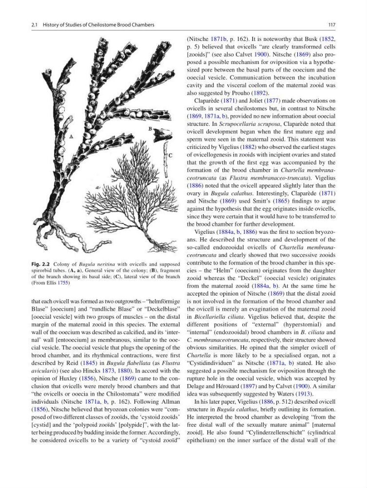
Fig. 2.2 Colony of Bugula neritina with ovicells and supposed
spirorbid tubes. (A, a), General view of the colony; ( B ), fragment
of the branch showing its basal side; ( C ), lateral view of the branch
(From Ellis 1755 )
2.1 History of Studies of Cheilostome Brood Chambers
that each ovicell was formed as two outgrowths – “helmförmige
Blase” [ooecium] and “rundliche Blase” or “Deckelblase”
[ooecial vesicle] with two groups of muscles – on the distal
margin of the maternal zooid in this species. The external
wall of the ooecium was described as calcifi ed, and its ‘internal’ wall [entooecium] as membranous, similar to the ooecial vesicle. The ooecial vesicle that plugs the opening of the
brood chamber, and its rhythmical con tractions, were fi rst
described by Reid ( 1845 ) in Bugula fl abellata (as Flustra
avicularis ) (see also Hincks 1873 , 1880 ). In accord with the
opinion of Huxley ( 1856 ), Nitsche ( 1869 ) came to the conclusion that ovicells were merely brood chambers and that
“the ovicells or ooecia in the Chilostomata” were modifi ed
individuals (Nitsche 1871a , b , p. 162). Following Allman
( 1856 ), Nitsche believed that bryozoan colonies were “composed of two different classes of zooïds, the ‘cystoid zooïds’
[cystid] and the ‘polypoid zooïds’ [polypide]”, with the latter being produced by budding inside the former. Accordingly,
he considered ovicells to be a variety of “cystoid zooïd”
(Nitsche 1871b , p. 162). It is noteworthy that Busk ( 1852 ,
p. 5) believed that ovicells “are clearly transformed cells
[zooids]” (see also Calvet 1900 ). Nitsche ( 1869 ) also proposed a possible mechanism for oviposition via a hypothesized pore between the basal parts of the ooecium and the
ooecial vesicle. Communication between the incubation
cavity and the visceral coelom of the maternal zooid was
also suggested by Prouho ( 1892 ).
Claparède ( 1871 ) and Joliet ( 1877 ) made observations on
ovicells in several cheilostomes but, in contrast to Nitsche
( 1869 , 1871a , b ), provided no new information about ooecial
structure. In Scrupocellaria scruposa , Claparède noted that
ovicell development began when the fi rst mature egg and
sperm were seen in the maternal zooid. This statement was
criticized by Vigelius ( 1882 ) who observed the earliest stages
of ovicellogenesis in zooids with incipient ovaries and stated
that the growth of the fi rst egg was accompanied by the
formation of the brood chamber in Chartella membranaceotruncata (as Flustra membranaceo-truncata ). Vigelius
( 1886 ) noted that the ovicell appeared slightly later than the
ovary in Bugula calathus . Interestingly, Claparède ( 1871 )
and Nitsche ( 1869 ) used Smitt’s ( 1865 ) fi ndings to argue
against the hypothesis that the egg originates inside ovicells,
since they were certain that it would have to be transferred to
the brood chamber for further development.
Vigelius ( 1884a , b , 1886 ) was the fi rst to section bryozoans. He described the structure and development of the
so-called endozooidal ovicells of Chartella membranaceotruncata and clearly showed that two successive zooids
contribute to the formation of the brood chamber in this species – the “Helm” (ooecium) originates from the daughter
zooid whereas the “Deckel” (ooecial vesicle) originates
from the maternal zooid ( 1884a , b ). At the same time he
accepted the opinion of Nitsche ( 1869 ) that the distal zooid
is not involved in the formation of the brood chamber and
the ovicell is merely an evagination of the maternal zooid
in Bicellariella ciliata . Vigelius believed that, despite the
different positions of “external” (hyperstomial) and
“internal” (endozooidal) brood chambers in B . ciliata and
C. membranaceotruncata , respectively, their structure showed
obvious similarities. He opined that the simpler ovicell of
Chartella is more likely to be a specialised organ, not a
“Cystidindividuen” as Nitsche ( 1871a , b ) stated. He also
suggested a possible mechanism for oviposition through the
rupture hole in the ooecial vesicle, which was accepted by
Delage and Hérouard ( 1897 ) and by Calvet ( 1900 ). A similar
idea was subsequently suggested by Waters ( 1913 ).
In his later paper, Vigelius ( 1886 , p. 512) described ovicell
structure in Bugula calathus , briefl y outlining its formation.
He interpreted the brood chamber as developing “from the
free distal wall of the sexually mature animal” [maternal
zooid]. He also found “Cylinderzellenschicht” (cylindrical
epithelium) on the inner surface of the distal wall of the
Fig. 2.2 Colony of Bugula neritina with ovicells and supposed
spirorbid tubes. (A, a), General view of the colony; ( B ), fragment
of the branch showing its basal side; ( C ), lateral view of the branch
(From Ellis 1755 )
2.1 History of Studies of Cheilostome Brood Chambers