

91
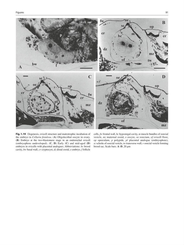
Fig. 1.19 Oogenesis, ovicell structure and matrotrophic incubation of
the embryo in Cellaria fi stulosa . ( A ) Oligolecithal oocyte in ovary.
( B ) Embryo at the two-blastomere stage in an endotoichal ovicell
(embryophore undeveloped). ( C , D ) Early ( C ) and mid-aged ( D )
embryos in ovicells with placental analogues. Abbreviations: bc brood
cavity, bw basal wall, cr cryptocyst, dz distal zooid, е embryo, f follicle
cells, fw frontal wall, hc hypostegal cavity, m muscle bundles of ooecial
vesicle, mz maternal zooid, o oocyte, ое ooecium, of ovicell floor,
op operculum, p polypide, pl placental analogue (embryophore),
sc sclerite of ooecial vesicle, tw transverse wall, v ooecial vesicle forming
brood sac. Scale bars: A – D , 20 μm
Figures
Fig. 1.19 Oogenesis, ovicell structure and matrotrophic incubation of
the embryo in Cellaria fi stulosa . ( A ) Oligolecithal oocyte in ovary.
( B ) Embryo at the two-blastomere stage in an endotoichal ovicell
(embryophore undeveloped). ( C , D ) Early ( C ) and mid-aged ( D )
embryos in ovicells with placental analogues. Abbreviations: bc brood
cavity, bw basal wall, cr cryptocyst, dz distal zooid, е embryo, f follicle
cells, fw frontal wall, hc hypostegal cavity, m muscle bundles of ooecial
vesicle, mz maternal zooid, o oocyte, ое ooecium, of ovicell floor,
op operculum, p polypide, pl placental analogue (embryophore),
sc sclerite of ooecial vesicle, tw transverse wall, v ooecial vesicle forming
brood sac. Scale bars: A – D , 20 μm
Figures