

90
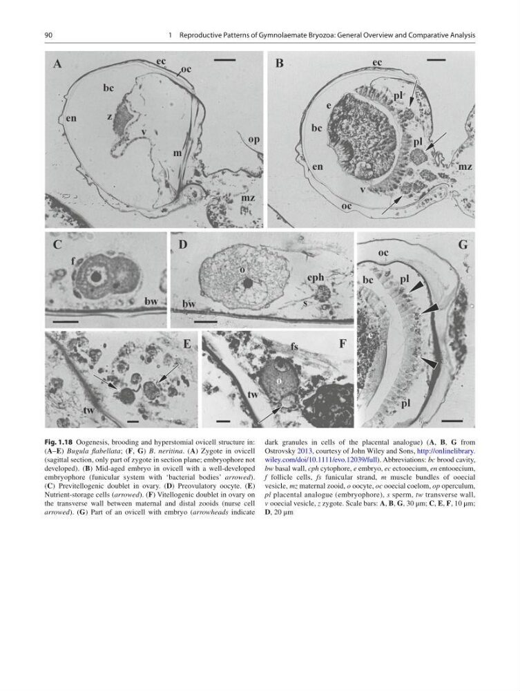
Fig. 1.18 Oogenesis, brooding and hyperstomial ovicell structure in:
( A – E ) Bugula fl abellata ; ( F , G ) B . neritina . ( A ) Zygote in ovicell
(sagittal section, only part of zygote in section plane; embryophore not
developed). ( B ) Mid-aged embryo in ovicell with a well-developed
embryophore (funicular system with ‘bacterial bodies’ arrowed ).
( C ) Previtellogenic doublet in ovary. ( D ) Preovulatory oocyte. ( E )
Nutrient-storage cells ( arrowed ). ( F ) Vitellogenic doublet in ovary on
the transverse wall between maternal and distal zooids (nurse cell
arrowed ). ( G ) Part of an ovicell with embryo ( arrowheads indicate
dark granules in cells of the placental analogue) ( A , B , G from
Ostrovsky 2013 , courtesy of John Wiley and Sons, http://onlinelibrary.
wiley.com/doi/10.1111/evo.12039/full ). Abbreviations: bc brood cavity,
bw basal wall, cph cytophore, е embryo, ес ectooecium, en entooecium,
f follicle cells, fs funicular strand, m muscle bundles of ooecial
vesicle, mz maternal zooid, o oocyte, oc ooecial coelom, op operculum,
pl placental analogue (embryophore), s sperm, tw transverse wall,
v ooecial vesicle, z zygote. Scale bars: A , B , G , 30 μm; C , E , F , 10 μm;
D , 20 μm
1 Reproductive Patterns of Gymnolaemate Bryozoa: General Overview and Comparative Analysis
Fig. 1.18 Oogenesis, brooding and hyperstomial ovicell structure in:
( A – E ) Bugula fl abellata ; ( F , G ) B . neritina . ( A ) Zygote in ovicell
(sagittal section, only part of zygote in section plane; embryophore not
developed). ( B ) Mid-aged embryo in ovicell with a well-developed
embryophore (funicular system with ‘bacterial bodies’ arrowed ).
( C ) Previtellogenic doublet in ovary. ( D ) Preovulatory oocyte. ( E )
Nutrient-storage cells ( arrowed ). ( F ) Vitellogenic doublet in ovary on
the transverse wall between maternal and distal zooids (nurse cell
arrowed ). ( G ) Part of an ovicell with embryo ( arrowheads indicate
dark granules in cells of the placental analogue) ( A , B , G from
Ostrovsky 2013 , courtesy of John Wiley and Sons, http://onlinelibrary.
wiley.com/doi/10.1111/evo.12039/full ). Abbreviations: bc brood cavity,
bw basal wall, cph cytophore, е embryo, ес ectooecium, en entooecium,
f follicle cells, fs funicular strand, m muscle bundles of ooecial
vesicle, mz maternal zooid, o oocyte, oc ooecial coelom, op operculum,
pl placental analogue (embryophore), s sperm, tw transverse wall,
v ooecial vesicle, z zygote. Scale bars: A , B , G , 30 μm; C , E , F , 10 μm;
D , 20 μm
1 Reproductive Patterns of Gymnolaemate Bryozoa: General Overview and Comparative Analysis