

83
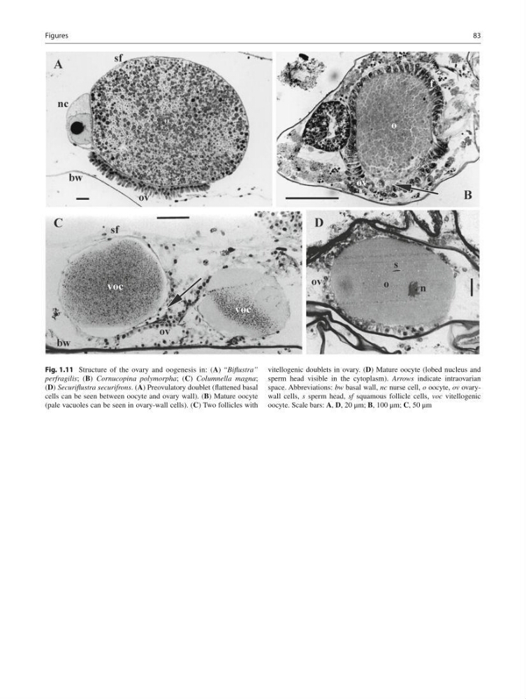
Fig. 1.11 Structure of the ovary and oogenesis in: ( A ) “ Bifl ustra”
perfragilis ; ( B ) Cornucopina polymorpha ; ( C ) Columnella magna ;
( D ) Securifl ustra securifrons . ( A ) Preovulatory doublet (fl attened basal
cells can be seen between oocyte and ovary wall). ( B ) Mature oocyte
(pale vacuoles can be seen in ovary-wall cells). ( C ) Two follicles with
vitellogenic doublets in ovary. ( D ) Mature oocyte (lobed nucleus and
sperm head visible in the cytoplasm). Arrows indicate intraovarian
space. Abbreviations: bw basal wall, nc nurse cell, o oocyte, ov ovarywall cells, s sperm head, sf squamous follicle cells, voc vitellogenic
oocyte. Scale bars: A , D , 20 μm; B , 100 μm; C , 50 μm
Figures
Fig. 1.11 Structure of the ovary and oogenesis in: ( A ) “ Bifl ustra”
perfragilis ; ( B ) Cornucopina polymorpha ; ( C ) Columnella magna ;
( D ) Securifl ustra securifrons . ( A ) Preovulatory doublet (fl attened basal
cells can be seen between oocyte and ovary wall). ( B ) Mature oocyte
(pale vacuoles can be seen in ovary-wall cells). ( C ) Two follicles with
vitellogenic doublets in ovary. ( D ) Mature oocyte (lobed nucleus and
sperm head visible in the cytoplasm). Arrows indicate intraovarian
space. Abbreviations: bw basal wall, nc nurse cell, o oocyte, ov ovarywall cells, s sperm head, sf squamous follicle cells, voc vitellogenic
oocyte. Scale bars: A , D , 20 μm; B , 100 μm; C , 50 μm
Figures