

82
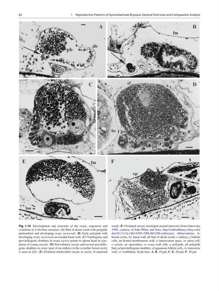
Fig. 1.10 Development and structure of the ovary, oogenesis and
ovulation in Cribrilina annulata . ( A ) Bud of distal zooid with polypide
primordium and developing ovary ( arrowed ). ( B ) Early polypide with
developing ovary ( arrowed ) on zooidal basal wall. ( C ) Vitellogenic and
previtellogenic doublets in ovary ( arrow points to sperm head in cytoplasm of young oocyte). ( D ) Preovulatory oocyte and several previtellogenic doublets in ovary (part of an embryo in the ovicellar brood cavity
is seen at left ). ( E ) Ovulated telolecithal oocyte in cavity of maternal
zooid. ( F ) Ovulated oocyte enveloped around introvert (from Ostrovsky
1998 , courtesy of John Wiley and Sons, http://onlinelibrary.wiley.com/
doi/10.1111/j.1463-6395.1998.tb01280.x/abstract ). Abbreviations: bc
brood cavity, bw basal wall, db bud of distal zooid, e embryo, f follicle
cells, fm frontal membranous wall, is intraovarian space, nc nurse cell,
o oocyte, op operculum, ov ovary-wall cells, p polypide, рb polypide
bud, pd previtellogenic doublets, sf squamous follicle cells, tw transverse
wall, ve vestibulum. Scale bars: A , B , 10 μm; C – E , 20 μm; F , 30 μm
1 Reproductive Patterns of Gymnolaemate Bryozoa: General Overview and Comparative Analysis
Fig. 1.10 Development and structure of the ovary, oogenesis and
ovulation in Cribrilina annulata . ( A ) Bud of distal zooid with polypide
primordium and developing ovary ( arrowed ). ( B ) Early polypide with
developing ovary ( arrowed ) on zooidal basal wall. ( C ) Vitellogenic and
previtellogenic doublets in ovary ( arrow points to sperm head in cytoplasm of young oocyte). ( D ) Preovulatory oocyte and several previtellogenic doublets in ovary (part of an embryo in the ovicellar brood cavity
is seen at left ). ( E ) Ovulated telolecithal oocyte in cavity of maternal
zooid. ( F ) Ovulated oocyte enveloped around introvert (from Ostrovsky
1998 , courtesy of John Wiley and Sons, http://onlinelibrary.wiley.com/
doi/10.1111/j.1463-6395.1998.tb01280.x/abstract ). Abbreviations: bc
brood cavity, bw basal wall, db bud of distal zooid, e embryo, f follicle
cells, fm frontal membranous wall, is intraovarian space, nc nurse cell,
o oocyte, op operculum, ov ovary-wall cells, p polypide, рb polypide
bud, pd previtellogenic doublets, sf squamous follicle cells, tw transverse
wall, ve vestibulum. Scale bars: A , B , 10 μm; C – E , 20 μm; F , 30 μm
1 Reproductive Patterns of Gymnolaemate Bryozoa: General Overview and Comparative Analysis