

490
13 The Structure and Metabolism of Carbohydrates
There are many examples illustrating the influence of the carbohydrate component on the conformation and physicochemical properties of glycoproteins. The carbohydrate portion of the integral membrane proteins is critical to their arrangement in the membrane and to their stability
[25,93]. The repulsion between molecules of the
negatively charged sialic acid-rich, 140-kDa glycoprotein podocalyxin on the podocytes of the
kidney glomeruli ensures that the filtration slits
between the interdigitating cell processes remain
open [65]. The sialic acids of the slime substances
(mucins) are responsible for their high viscosity
and lubrication properties; the high density of
charges forces the molecule into a stretched rodshaped form and with many carbohydrate chains
attached it resembles a bottle brush [2, 25]. The
slime substances secreted by fish epidermis greatly
reduce friction, even in dilute solutions with mucin
concentrations below 0.1 %. Thus, swimming fish
are constantly enveloped in a thin film of slime
which helps them to maintain considerably higher
velocities. Unfortunately, little is known about the
molecular structure of fish mucin. Glycoproteins
are important components of the jelly substances
enveloping the eggs of many vertebrates from the
fish to the birds [77, 131]. The gel-like consistency
of the albumen of birds is due to the cross-linking
of the glycoproteins ovomucin and lysozyme
(p.388). In the antifreeze glycoproteins in the
blood plasma of antarctic fish, it is the disaccharides lying on the surface which, by an unknown
mechanism, prevent the formation of ice crystals
(see Fig. 5.3, p. 207). The mechanical properties
of connective tissues are also determined by interaction of their protein and carbohydrate components with water and ions (p. 482).
a)
GalNAc ~Gal ~ GalNAcaThrlSer
t al,2 f a2,6
Fuc
NeuAc
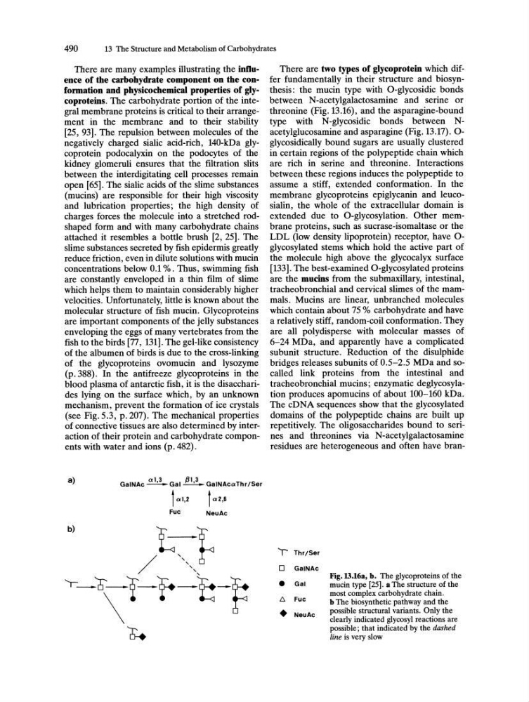
There are two types of glycoprotein which differ fundamentally in their structure and biosynthesis: the mucin type with O-glycosidic bonds
between N-acetylgalactosamine and serine or
threonine (Fig. 13.16), and the asparagine-bound
type with N-glycosidic bonds between Nacetylglucosamine and asparagine (Fig. 13.17).0glycosidically bound sugars are usually clustered
in certain regions of the polypeptide chain which
are rich in serine and threonine. Interactions
between these regions induces the polypeptide to
assume a stiff, extended conformation. In the
membrane glycoproteins epiglycanin and leucosialin, the whole of the extracellular domain is
extended due to O-glycosylation. Other membrane proteins, such as sucrase-isomaltase or the
LDL (low density lipoprotein) receptor, have 0glycosylated stems which hold the active part of
the molecule high above the glycocalyx surface
[133]. The best-examined O-glycosylated proteins
are the mucins from the submaxillary, intestinal,
tracheobronchial and cervical slimes of the mammals. Mucins are linear, unbranched molecules
which contain about 75 % carbohydrate and have
a relatively stiff, random-coil conformation. They
are all polydisperse with molecular masses of
6-24 MDa, and apparently have a complicated
subunit structure. Reduction of the disulphide
bridges releases subunits of 0.5-2.5 MDa and socalled link proteins from the intestinal and
tracheobronchial mucins; enzymatic deglycosylation produces apomucins of about 100-160 kDa.
The cDNA sequences show that the glycosylated
domains of the polypeptide chains are built up
repetitively. The oligosaccharides bound to serines and threonines via N-acetylgalactosamine
residues are heterogeneous and often have branT ThrlSer
o GalNAc
•
Gal
1:,.
Fuc
•
NeuAc
Fig. 13.16a, b. The glycoproteins of the
mucin type [25]. a The structure of the
most complex carbohydrate chain.
b The biosynthetic pathway and the
possible structural variants. Only the
clearly indicated glycosyl reactions are
possible; that indicated by the dashed
line is very slow