

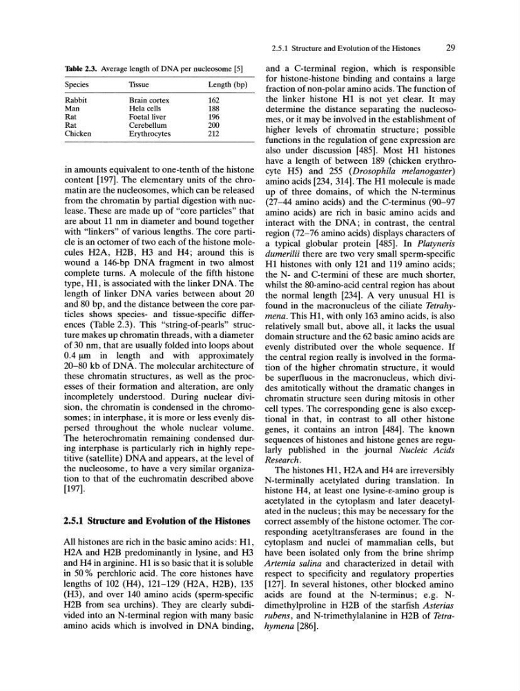
Table 2.3. Average length of DNA per nucleosome [5]
Species
Tissue
Length (bp)
Rabbit
Brain cortex
162
Man
Hela cells
188
Rat
Foetal liver
196
Rat
Cerebellum
200
Chicken
Erythrocytes
212
in amounts equivalent to one-tenth of the histone
content [197]. The elementary units of the chromatin are the nucleosomes, which can be released
from the chromatin by partial digestion with nuclease. These are made up of "core particles" that
are about 11 nm in diameter and bound together
with "linkers" of various lengths. The core particle is an octomer of two each of the histone molecules H2A, H2B, H3 and H4; around this is
wound a 146-bp DNA fragment in two almost
complete turns. A molecule of the fifth histone
type, H1, is associated with the linker DNA. The
length of linker DNA varies between about 20
and 80 bp, and the distance between the core particles shows species- and tissue-specific differences (Table 2.3). This "string-of-pearls" structure makes up chromatin threads, with a diameter
of 30 nm, that are usually folded into loops about
O.4!-lm in length and with approximately
20-80 kb of DNA. The molecular architecture of
these chromatin structures, as well as the processes of their formation and alteration, are only
incompletely understood. During nuclear division, the chromatin is condensed in the chromosomes; in interphase, it is more or less evenly dispersed throughout the whole nuclear volume.
The heterochromatin remaining condensed during interphase is particularly rich in highly repetitive (satellite) DNA and appears, at the level of
the nucleosome, to have a very similar organization to that of the euchromatin described above
[197].
2.5.1 Structure and Evolution of the Histones
All histones are rich in the basic amino acids: H1,
H2A and H2B predominantly in lysine, and H3
and H4 in arginine. H1 is so basic that it is soluble
in 50 % perchloric acid. The core histones have
lengths of 102 (H4), 121-129 (H2A, H2B) , 135
(H3), and over 140 amino acids (sperm-specific
H2B from sea urchins). They are clearly subdivided into an N-terminal region with many basic
amino acids which is involved in DNA binding,
2.5.1 Structure and Evolution of the Histones
29
and a C-terminal region, which is responsible
for histone-histone binding and contains a large
fraction of non-polar amino acids. The function of
the linker histone H1 is not yet clear. It may
determine the distance separating the nucleosomes, or it may be involved in the establishment of
higher levels of chromatin structure; possible
functions in the regulation of gene expression are
also under discussion [485]. Most H1 his tones
have a length of between 189 (chicken erythrocyte H5) and 255 (Drosophila melanogaster)
amino acids [234, 314]. The H1 molecule is made
up of three domains, of which the N-terminus
(27-44 amino acids) and the C-terminus (90-97
amino acids) are rich in basic amino acids and
interact with the DNA; in contrast, the central
region (72-76 amino acids) displays characters of
a typical globular protein [485]. In Platyneris
dumerilii there are two very small sperm-specific
H1 histones with only 121 and 119 amino acids;
the N- and C-termini of these are much shorter,
whilst the 80-amino-acid central region has about
the normal length [234]. A very unusual H1 is
found in the macronucleus of the ciliate Tetrahymena. This H1, with only 163 amino acids, is also
relatively small but, above all, it lacks the usual
domain structure and the 62 basic amino acids are
evenly distributed over the whole sequence. If
the central region really is involved in the formation of the higher chromatin structure, it would
be superfluous in the macronucleus, which divides amitotically without the dramatic changes in
chromatin structure seen during mitosis in other
cell types. The corresponding gene is also exceptional in that, in contrast to all other histone
genes, it contains an intron [484]. The known
sequences of his tones and histone genes are regularly published in the journal Nucleic Acids
Research.
The histones H1, H2A and H4 are irreversibly
N-terminally acetylated during translation. In
histone H4, at least one lysine-E-amino group is
acetylated in the cytoplasm and later deacetylated in the nucleus; this may be necessary for the
correct assembly of the histone octomer. The corresponding acetyltransferases are found in the
cytoplasm and nuclei of mammalian cells, but
have been isolated only from the brine shrimp
Artemia salina and characterized in detail with
respect to specificity and regulatory properties
[127]. In several histones, other blocked amino
acids are found at the N-terminus; e.g. Ndimethylproline in H2B of the starfish Asterias
rubens, and N-trimethylalanine in H2B of Tetrahymena [286].
Species
Tissue
Length (bp)
Rabbit
Brain cortex
162
Man
Hela cells
188
Rat
Foetal liver
196
Rat
Cerebellum
200
Chicken
Erythrocytes
212
in amounts equivalent to one-tenth of the histone
content [197]. The elementary units of the chromatin are the nucleosomes, which can be released
from the chromatin by partial digestion with nuclease. These are made up of "core particles" that
are about 11 nm in diameter and bound together
with "linkers" of various lengths. The core particle is an octomer of two each of the histone molecules H2A, H2B, H3 and H4; around this is
wound a 146-bp DNA fragment in two almost
complete turns. A molecule of the fifth histone
type, H1, is associated with the linker DNA. The
length of linker DNA varies between about 20
and 80 bp, and the distance between the core particles shows species- and tissue-specific differences (Table 2.3). This "string-of-pearls" structure makes up chromatin threads, with a diameter
of 30 nm, that are usually folded into loops about
O.4!-lm in length and with approximately
20-80 kb of DNA. The molecular architecture of
these chromatin structures, as well as the processes of their formation and alteration, are only
incompletely understood. During nuclear division, the chromatin is condensed in the chromosomes; in interphase, it is more or less evenly dispersed throughout the whole nuclear volume.
The heterochromatin remaining condensed during interphase is particularly rich in highly repetitive (satellite) DNA and appears, at the level of
the nucleosome, to have a very similar organization to that of the euchromatin described above
[197].
2.5.1 Structure and Evolution of the Histones
All histones are rich in the basic amino acids: H1,
H2A and H2B predominantly in lysine, and H3
and H4 in arginine. H1 is so basic that it is soluble
in 50 % perchloric acid. The core histones have
lengths of 102 (H4), 121-129 (H2A, H2B) , 135
(H3), and over 140 amino acids (sperm-specific
H2B from sea urchins). They are clearly subdivided into an N-terminal region with many basic
amino acids which is involved in DNA binding,
2.5.1 Structure and Evolution of the Histones
29
and a C-terminal region, which is responsible
for histone-histone binding and contains a large
fraction of non-polar amino acids. The function of
the linker histone H1 is not yet clear. It may
determine the distance separating the nucleosomes, or it may be involved in the establishment of
higher levels of chromatin structure; possible
functions in the regulation of gene expression are
also under discussion [485]. Most H1 his tones
have a length of between 189 (chicken erythrocyte H5) and 255 (Drosophila melanogaster)
amino acids [234, 314]. The H1 molecule is made
up of three domains, of which the N-terminus
(27-44 amino acids) and the C-terminus (90-97
amino acids) are rich in basic amino acids and
interact with the DNA; in contrast, the central
region (72-76 amino acids) displays characters of
a typical globular protein [485]. In Platyneris
dumerilii there are two very small sperm-specific
H1 histones with only 121 and 119 amino acids;
the N- and C-termini of these are much shorter,
whilst the 80-amino-acid central region has about
the normal length [234]. A very unusual H1 is
found in the macronucleus of the ciliate Tetrahymena. This H1, with only 163 amino acids, is also
relatively small but, above all, it lacks the usual
domain structure and the 62 basic amino acids are
evenly distributed over the whole sequence. If
the central region really is involved in the formation of the higher chromatin structure, it would
be superfluous in the macronucleus, which divides amitotically without the dramatic changes in
chromatin structure seen during mitosis in other
cell types. The corresponding gene is also exceptional in that, in contrast to all other histone
genes, it contains an intron [484]. The known
sequences of his tones and histone genes are regularly published in the journal Nucleic Acids
Research.
The histones H1, H2A and H4 are irreversibly
N-terminally acetylated during translation. In
histone H4, at least one lysine-E-amino group is
acetylated in the cytoplasm and later deacetylated in the nucleus; this may be necessary for the
correct assembly of the histone octomer. The corresponding acetyltransferases are found in the
cytoplasm and nuclei of mammalian cells, but
have been isolated only from the brine shrimp
Artemia salina and characterized in detail with
respect to specificity and regulatory properties
[127]. In several histones, other blocked amino
acids are found at the N-terminus; e.g. Ndimethylproline in H2B of the starfish Asterias
rubens, and N-trimethylalanine in H2B of Tetrahymena [286].